
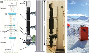
 Schematic and images of the common-mode digital holographic microscope in its laboratory and field implementation. (a) Schematic of digital holographic microscope, showing four main elements: source, sample, microscope optics and sensor. (b) Solid model and (c) instrument in the laboratory. (d) Field work of the instrument involved measuring bacteria in the sea ice of Greenland.
Bacterial motility plays a role in critical environmental and physiological processes, including nutrient cycling, biofouling and virulence. Despite its importance, motility has been studied in only a few test organisms because of the difficulties of imaging moving micrometer-sized cells. However, the study of the motility of microorganisms is a field that promises to be revolutionized by digital holographic microscopy (DHM).1
Advances in high-speed computers and large-format digital imagers have led to a burgeoning interest in DHM. In DHM, recording of the optical interference—fringes—is is done with an array detector.2,3 Recording fringes of high contrast over the detector integration time requires good static optical coherence and fringe stability during the exposure time.4
Digital holography adds two unique capabilities over classic holography: the ability to numerically reconstruct the electric field within a volume and to do so as a function of time. Thus it is possible to create a time-lapse movie of the electric field within a three-dimensional volume. Imaging moving bacteria is challenging because of the small size of the cells, their rapid motion (tens to hundreds of cell lengths per second), and their low intensity contrast. We have developed a common-mode instrument that has sufficient spatial and temporal resolution to enable these measurements, including quantitative phase contrast.5
Typical designs for off-axis systems result in rather large instruments (usually Mach–Zehnder or Michelson-type), which are alignment-sensitive and generally less suited for extreme environments. Our new design maintains the off-axis implementation but with a robust optical design that does not sacrifice performance. This optical design avoids the limitations of prior systems and provides many advantages, including compactness, intrinsic stability, robustness against misalignment, ease of assembly, and cost. Moreover, it supports use in both routine laboratory settings as well as extreme environments without any sacrifice in performance, enabling ready observation of microbial species in the field.
Researchers
J. Kent Wallace, Eugene Serabyn, Jonas Kühn, Kurt Liewer and Chris Lindensmith, Jet Propulsion Laboratory, Pasadena, Calif., USA
Stephanie Rider, California Institute of Technology, Pasadena, Calif., USA
Jody Deming and Gordon Showalter, University of Washington, Seattle, Wash., USA
Jay Nadeau, McGill University, Montreal, Quebec, Canada, and California Institute of Technology
References
1. C. Knox. Science 153, 989 (1966).
2. U. Schnars and W. Jüptner. Meas. Sci. Technol. 13, R85 (2002).
3. E. Cuche et al. Appl. Opt. 38, 6994 (1999).
4. E. Cuche et al. Appl. Opt. 39, 4070 (2000).
5. J.K. Wallace et al. Opt. Express 23, 17367 (2015).