
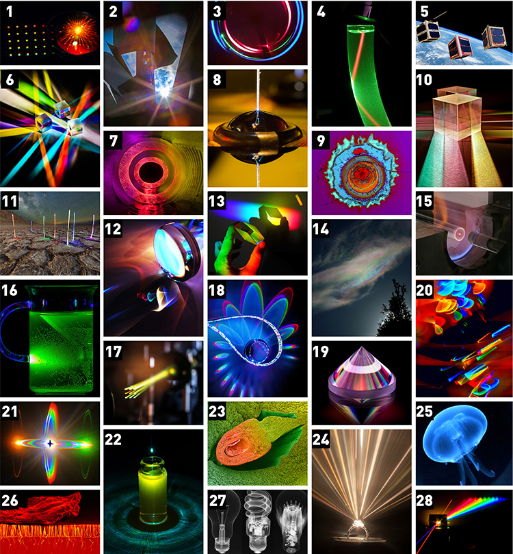
1. A droplet of fluorescent molecules sits atop an array of nano-sized holes. An excitation laser from below is diffracted by the nanohole array, causing a "bloom" of internally reflected fluorescence within the droplet. The fluorescence from this array was used to calibrate the field-dependent aberrations of a super-resolved fluorescence microscope (see article submitted to Optica, 2015). Winner 2015 After Image photo contest. [Credit: Matthew D. Lew, Washington University in St. Louis, USA]
2. A view of the 21 August solar eclipse through the arms of The Sentinel, a large metal sculpture by Albert Paley at the Rochester Institute of Technology (RIT). A tight camera aperture (f/32) creates a starburst image, while reflections within the camera system cause colorful ghost images of the partially eclipsed sun. [Credit: Grover Swartzlander, RIT, USA]
3. Laser scattering in a glass cylinder, the light rays split and bounce back and forth , making the long term prediction of the trajectories of these rays very difficult. Honorable mention 2018 After Image photo contest. [Credit: Albetro Tufaile, Soft Matter Lab, EACH-USP]
4. A green laser beam propagating in a plastic tube filled with olive oil shows total internal reflection and the Goos-Hänchen shift on dielectric boundaries. Reddish color of the beam inside
the oil is due to the fluorescence of chlorophyll molecules. [Credit: Emilio Gómez-González, Universidad de Sevilla, Spain]
5. CubeSats with Earth in the background. [Credit: NASA]
6. RGB combiner cubes are used in projection systems to combine the red, green and blue components of an image. These same cubes can act as vibrant color splitters for incident light. Winner 2014 After Image photo contest. [Credit: Page E. King, College of Optical Sciences, Tucson, Ariz., USA]
7. Spirographs created in a dark tunnel through the art of light painting. [Credit: Johnny A. Andrews, http://stimulightthenight.com]
8. An optical fibre with 3D printed doublet Fresnel lenses is inserted into a water droplet to image the beam profile by an inverse microscope. A white light laser passed through the objective is used to align the fibre in respect to the objective and causes the blue colour of the lenses in the refracted image on the surface of the droplet. The image is taken by a 100mm f/2.8 micro lens on a DSLR (Canon 60D). The image is cropped, and gone under noise reduction and contrast enhancement. This photo involves equal contribution of two colleagues: measurement setup and fabrication by Dr Asa Asadollahbaik, photography by Mr Moritz Floess. The fabrication and design also includes the collaboration of Mr Simon Thiele and Ms Ksenia Weber. The image is taken at Optics Lab in 4th Physics Institute (Head of the Institute: Professor Harald Giessen), University of Stuttgart, Stuttgart. [Credit: Asa Asadollahbaik, 4th Physics Institute, University of Stuttgart]
9. Microscopic image of damage caused by intense laser irradiation on a dielectric surface. Honorable mention, 2019 After Image photo contest. [Credit: LIDARIS Laser Damage Testing Team, Vilnius, Lithuania]
10. White light strikes a pair of beamsplitter cubes with anti-reflective coatings for infrared light. These coatings separate white light into its constituent colors. The surrounding lab environment was darkened to highlight the mesmerizing colors captured in this picture with a cellphone camera. The texture of the sheet of paper on which the cubes were placed can be seen on the front of the image. Second place winner 2019 After Image photo contest. [Credit: Gibraham A. Graciano, CICESE, Mexico]
11. Nighttime view over Badwater, Death Valley, Calif., USA, the lowest point (-282 feet below sea level) in the United States. The “aliens” are actually made from combined exposures of LED lights waved in a more-or-less vertical direction. The base picture is a 15-second exposure with the core of the Milky Way galaxy in view. [Credit: Michael Duncan, Optica Senior Science Advisor]
12. Holiday lights imaged by a lens. OPN 2013 After Image Photo Contest. [Credit: Page E. King, College of Optical Sciences, Tucson, Ariz., USA]
13. Dispersing Rhombus. [Credit: Karen L. Bridges]
14. Iridescent cloud over North Bend, Oregon, Nikon Sure Shot Camera. [Credit: Robert Schalck, Senior Member]
15. Rare earth doped fibres starts with the preform fabrication. In Modified Chemical Vapour Deposition technique, select chemicals are fused and deposited layer by layer onto a glass tube that rotates on a lathe after the chemicals are subject to fire. The pictures was taken in the Optoelectronics Research Centre at the University of Southampton with a Huawei P9 mobile with no filter. OPN 2017 After Image Photo Contest. [Credit: Norberto Ramirez, University of Southampton, UK]
16. A blue/UV laser pointer was coupled into the handle of a cup, which guides it and feeds it into a fluorescing liquid that lights up in green colors. Surrounding Lab environment was darkened completely to highlight the effect. Mild post-processing was done (clipping, lifting the contrasts, noise correction). The image was taken in the lab at Leibniz-IPHT Jena. Winner 2018 After Image photo contest. [Credit: Tobias Tieß, Leibniz-IPHT]
17. A spatial light modulator (SLM) is used to generate nine supercontinuum beams. The flexibility of the SLMs allows for a control of the intensity and spectral content of the generated light.
[Credit: Rocio Borrego Varillas and Jorge Pérez Vizcaíno, Universitat Jaume I, Spain]
18. A strip of LEDs creates white light by combing sets of red, green and blue LEDs placed together. When imaged through the glass sphere in the center, the red, green and blue caustics are imaged to slightly different locations, due to the small distances between each color LED. Second Place 2016 After Image photo contest. [Credit: Page E. King, College of Optical Sciences, University of Arizona, USA]
19. A solid glass Fresnel cone, illuminated by a rainbow-colored light source. The cone uses total internal reflection of incident polarized light to easily generate white-light orbital angular momentum and radially polarized vector vortex beams. Image taken in collaboration with QuantIC imaging hub, based at the University of Glasgow, U.K. Honorable mention, 2019 After Image photo contest. [Credit: Kevin J. Mitchell, University of Glasgow]
20. String of lights imaged onto a detector plane by a misaligned singlet lens. [Credit: Page E. King, College of Optical Sciences, Tucson, AZ, USA]
21. Acoustic waves spectrally modulate supercontinuum light inside an acousto-optic tunable filter. The supercontinuum light interacts with resonating acoustic modes to produce 4-lobed orange, green and rainbow Schaefer-Bergmann diffraction patterns around the two spectrally modulated beams and undiffracted light. These optical patterns can quantitatively measure acoustic properties of crystals. Winner 2016 After Image photo contest. [Credit: Stephanie Swartz, University of Colorado at Boulder, USA]
22. Output light field of a tapered fiber probe illuminating a vial filled with water containing trace amounts of the dye Rhodamine 6B. Coupled into the fiber are green (532 nm) and blue (488 nm) laser light under slightly different angles resulting in colorful patterns due to effects of diffraction, interference, scattering and fluorescence. The result is reminiscent of a human iris with the pupil staring intently at the bottom of the glass vial from close proximity. [Credit: Nils Frederik Hasselmann and Mathias Hünecke, Nonlinear Photonics, Münster University, Germany]
23. The picture was shot at Offenburg University using a scanning electron microscope (SEM) by Pascale Müller, and then digitally mastered by Dan Curticapean.
First-place winner OPN 2019 After Image Photo Contest [Credit: Dan Curticapean, Offenburg University, Germany]
24. The sparkles from my wife’s engagement ring are visualized by a collimated supercontinuum laser incident normal to the largest diamond in the ring. A piece of paper is used to trace out the beam in this long exposure image. Small rainbow beams can be seen shooting away from the stone, proof that the dispersive design of the diamond is doing its job. Second place winner, 2018 After Image photo contest. [Credit: Benjamin Cromey, University of Arizona]
25. Translucent body of a Moon jellyfish Charleston Marine Life Center, Charleston, OR, USA. [Credit: Robert Schalck]
26. The image shows a chunk of dust on a silicon substrate with nanowire-like structure on the the surface, which resembles a meteorite falling on a forest. The surface is coated with chromium for better resolution in SEM. Third place winner (tie), 2018 After Image photo contest. [Credit: Bingtao Gao, Univeristy of Iowa, USA]
27. Imaged with the X-rays of Nikon's XTH225 CT system [Credit: Herminso Villarraga-Gómez, Nikon Metrology Americas & University of North Carolina at Charlotte, USA]
28. Generation of Raman sidebands in diamond. Two Gaussian laser beams, pump (purple) and Stokes (dark orange), enter a Raman medium (diamond) and generate a broadband of higher order anti-Stokes. The photo was taken with a Canon EOS 80D, by moving a white screen in front of the beams at a 30-second time exposure. Honorable mention, 2019 After Image photo contest. [Credit: Aysan Bahari, Texas A&M University]
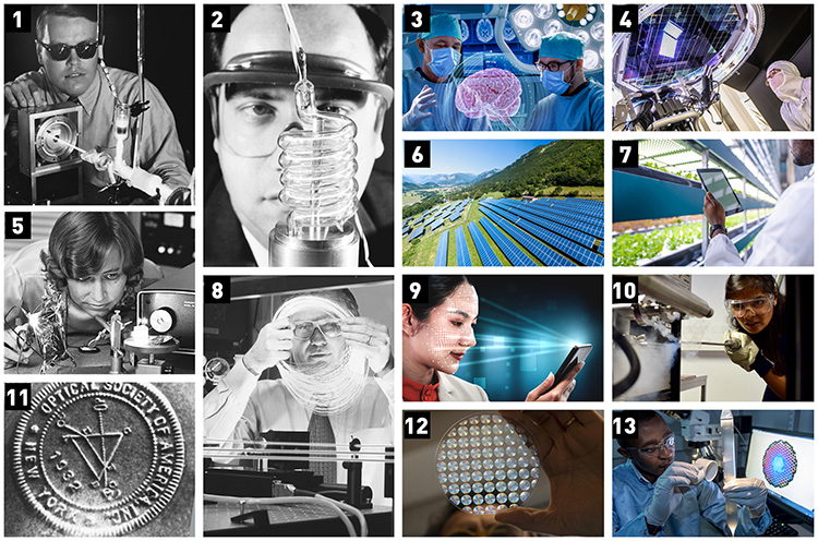
1. Future OSA Fellow William Silfvast with an early He-Cd laser at Bell Labs in 1969. [Credit: Alcatel-Lucent U.S.A., Inc.]
2. Theodore Maiman, 1960, pictured with an early ruby laser flashlamp assembly. [Credit: Courtesy of HRL Laboratories]
3. Credit: Getty Images
4. The large CCD array at the heart of the Vera C. Rubin Observatory’s ultrasensitive camera. [Credit: J.Orrell/SLAC National Accelerator Laboratory]
5. Elsa Garmire, a future OSA President and a pioneer in nonlinear optics, in the lab at the California Institute of Technology, circa 1969. [Courtesy of the Archives, California Institute of Technology]
6. Credit: Getty Images
7. Credit: Getty Images
8. Optica Fellow David N. Payne of the University of Southampton, U.K., was instrumental in developing erbium-doped fiber amplifiers, which helped enable modern fiber communications. [Credit: ORC, University of Southampton]
9. Credit: Getty Images
10. Author Himani Arora in the materials characterization lab. [Credit: Courtesy of H. Arora]
11. OSA’s seal of incorporation, 1932. [Optica, formerly OSA]
12. Giuseppe Strangi views a metalens array. [Credit: Giuseppe Strangi & Federico Capasso]
13. Researcher Eric Numkam Fokoua checks a microstructured fiber preform in the Optoelectronics Research Center at the University of Southampton, U.K. [Credit: University of Southampton]
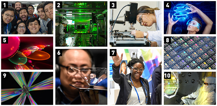
1. Credit: Optica, formerly OSA
2. Researchers created high-intensity pulses using the petawatt laser (pictured) at the Center for Relativistic Laser Science (CoReLS) in the Republic of Korea. [Image: Chang Hee Nam, CoReLS]
3. Credit: Getty Images
4. Credit: Getty Images
5. Caustics formed by LEDs imaging through a glass sphere. OPN 2013 After Image Photo Contest. [Credit: Page E. King, College of Optical Sciences, Tucson, Ariz., USA]
6. Credit: Optica, formerly OSA
7. Credit: Optica, formerly OSA
8. An array on a scrap silicon wafer. [Credit: Page E. King, College of Optical Sciences, Tucson, Ariz., USA]
9. An arrangement of plastic flutes between a linear- and a circular-polarizer. The colors represent wavelength-dependent retardance paths that rotate the polarization orientation through different angles. Honorable mention, 2018 After Image photo contest. [Credit: Samuel F. Pellicori, Senior Member OSA]
10. A LIGO technician examines one of the facility’s mirrors. A research team recently used precise measurements enabled by the LIGO lasers and mirrors to cool a 10-kg object to near its quantum ground state of motion. [Credit: Matt Heintze/Caltech/MIT/LIGO Lab]