
Scatterings
Scientists Image Rods in the Living Eye
New research demonstrates a way to image tiny but important light receptors in the living eye.
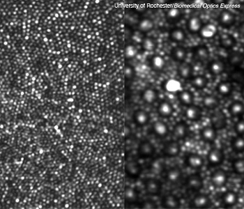 (Left) The smallest cones at the center of the retina (the fovea). (Right) The large bright dots with a dark ring around them are cones, and the surrounding (and far more abundant) smaller spots are rods.
Nearly 95 percent of the photoreceptors in our eyes are rods, the 2-µm-diameter cells that sense low levels of light. New research at the University of Rochester (N.Y., U.S.A.) demonstrates a way to image these tiny but important receptors in the living eye (Biomed. Opt. Express 2, 1864, 2011; doi:10.1364/BOE.2.001864).
…Log in or become a member to view the full text of this article.
This article may be available for purchase via the search at Optica Publishing Group.
Optica Members get the full text of Optics & Photonics News, plus a variety of other member benefits.