
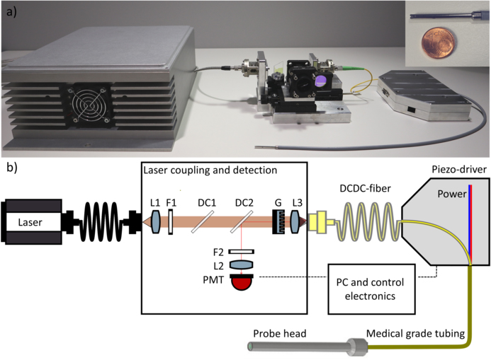
Photo and schematic of the fiber probe and laser coupling unit. [Image: E. Pshenay-Severin et al.] [Enlarge image]
A custom-designed fiber optic cable reportedly may help surgeons better map and remove cancerous tumors (Light Sci. Appl., doi: 10.1038/s41377-021-00648-w). Employed in a new endoscope probe, the fiber allows advanced in-body imaging that, according to the authors, could help doctors differentiate between healthy and malignant tissue by showing both morphological and biochemical information.
The researchers believe that the new endoscope could improve patient care and help reduce expensive follow-up diagnostics. In its current state, the endoscope is nearly ready for testing in a medical environment. Eventually, after such testing and clinical approval, the team believes it could even be used during surgery.
“The reported endoscope concept now marks a step forward to identify tumor tissue in real-time during surgery without cutting,” said author Bernhard Messerschmidt, with GRINTECH, Jena, Germany. “The fiber probe opens new possibilities for improved intraoperative disease diagnostics directly linked to better patient care.”
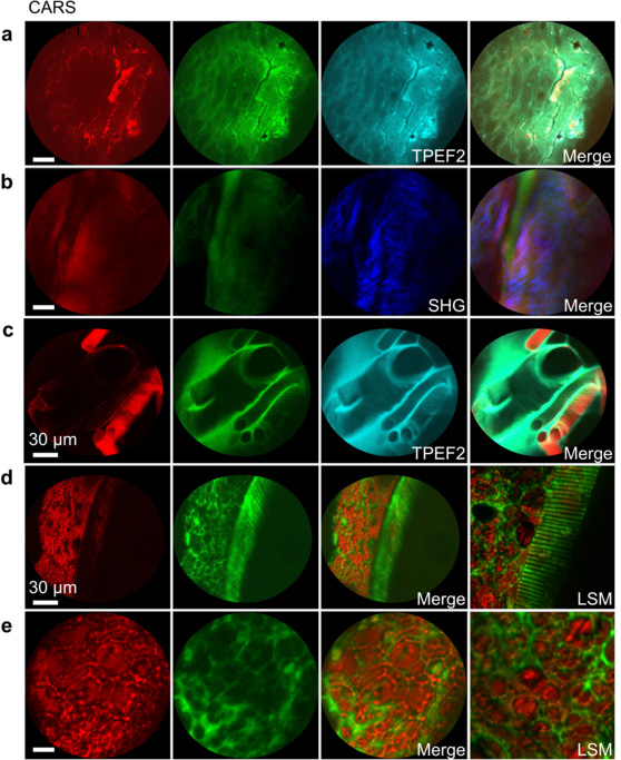
Multimodal CARS/SHG/TPEF endoscopic images of unstained biological samples from the new probe, showing CARS (first column), TPEF1 (second column), additional imaging modalities as labeled (third column), and merged data (fourth column). Samples include human epithelial tissue from head and neck (a), dura mater section of ovis aries rich in collagen (b), and sections of Galleria mellonella larvae (c, d, e). [Image: E. Pshenay-Severin et al.] [Enlarge image]
The good, the bad, and the invisible
Cancer can be hard to eliminate from a body, as malignant tumors often lurk amid healthy tissue. When removing a cancerous tumor, a surgeon must be careful to extract as much of the tumor as possible—if it isn’t fully removed, it can grow back like a weed.
Previously, researchers have found that malignant tumors can be identified with the combination of three types of imaging—coherent anti-Stokes Raman scattering (CARS), second-harmonic generation (SHG) and two-photon excited fluorescence (TPEF). But this type of multimodal nonlinear endoscopy has been extremely difficult to do in the human body, because of both size requirements and the fact that important properties of the pulsed laser light are lost the transport in fibers. Instead, tissues are typically removed by biopsy and inspected under a microscope after staining—a multi-day process.
Separate, yet together
To enable in-body imaging, a team of researchers from GRINTECH and the Leibniz Institute of Photonic Technology, Jena, led by Optica Senior Member Juergen Popp, developed a new platform that combines advances in fiber optic cables, a special laser source and custom optics. The resulting endoscope has sub-cellular resolution and a 180-µm field of view, packaged into an ultra-compact platform—the laser source and optical units are each the size of a shoebox and the probe tip is smaller than a pen.
A key advance in the new endoscope is a custom designed pure silica fiber optic cable with a double core, produced at the Leibniz Institute with a stack-and-draw technique. The solid, single-mode, double-clad fiber allows different laser signals to be guided separately without interfering each other. At the tip of the endoscope, a linear diffractive optical grating overlays the signals to allow full field of view, high-resolution imaging of the tissue.