
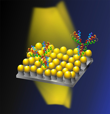
OIST researchers have devised a biosensor surface covered with millions of gold-capped “nanomushrooms,” which can monitor cell growth in real time by reading changes in refractive index as cells proliferate on the surface. [Image: OIST]
Medical researchers need ways to monitor, over long timescales, the proliferation of live cells used in laboratory drug tests and other clinical studies. But many assays for that purpose require complex labeling of cells with outside agents that hinder real-time monitoring over long periods. Now, a research team at the Okinawa Institute of Science and Technology (OIST) Graduate University, Japan, has created a label-free plasmonic biosensing material that can keep track of cell growth for as long as seven days—using an array of optical antennas shaped like tiny mushrooms (Adv. Biosyst., doi: 10.1002/adbi.201700258).
Problematic assays
Most assays used to measure cell proliferation rely on dyeing, staining or labeling cells to allow their activity to be monitored using fluorescence, colorimetry or other techniques. Some even involve tagging cells with a radioactive marker. These techniques can be costly and labor-intensive. Further, over the longer timescales required for some lab studies, which can extend across days, the external labels can themselves interfere with the cell behaviors being monitored, or even lead to cell death.
Label-free optical techniques, such as plasmonic biosensors, can avoid some of these cost and complexity drawbacks. But it’s been tough to come up with a nanoplasmonic substrate with a large surface area on which cells can survive over the long haul.
“Mushrooms” of glass and gold
The OIST team, led by Amy Shen of the university’s Micro/Bio/Nanofluidics unit, wanted to come up with a nanosensor geometry on which cells could survive and grow for a long period, without sensor-induced changes in their behavior. To get there, they began with an ordinary glass slide, on which they deposited a 4-nm-thick gold film. Thermal dewetting of the film at 560 °C led to the formation of tiny gold blobs or islands on the slide. Finally, a reactive-ion etching step, using an SF6 plasma, selectively peeled away the glass substrate around the nanoblobs.
The result was a field of mushroom-shaped nanostructures, each roughly 20 nm in width, consisting of 20-nm-thick gold caps atop glass stems 30 to 40 nm tall and distributed roughly 10 nm from one another across the slide. The subwavelength concentration and amplification of light shone on the gold structures causes local surface plasmon resonances; those resonances themselves are enhanced by the mushroom structure, which boosts the 3-D field strength around the gold mushroom caps.
The sensor works by detecting changes in refractive index as cells proliferate and bind to the nanostructure surface. In the setup, light is shone through the top of the nanosurface on which a cell-containing solution has been deposited, and the transmitted light is picked up and spectroscopically analyzed at the bottom. As cells proliferate and adhere to the nanomushrooms with membrane proteins, the process leads to changes in the peak wavelength of the plasmonic resonance. Those changes, read in the transmitted light, can in turn be mapped to refractive-index changes, to give a reading on cell growth in real time.
Seven-day experiment
The OIST team tested the system using live fibroblast cells, in an experiment running seven days. Comparing the nanosurface’s results with visual estimates using a cell-counting tool known as a hemocytometer, the researchers found that the nanomushrooms were sensitive enough to reliably pick up, in the first hour and a half of the experiment, an increase of 160 cells within an initial population of 10,000 cells. And the system was able to continue counting the proliferating fibroblasts across the entire weeklong experiment, with the nanostructures causing no apparent harm to cell growth.
The OIST researchers believe that, because it can provide real-time information on cell-growth kinetics, the nanosurface they’ve designed complements other cell-counting techniques and assays, which provide only limited information on that head. The team also thinks the nanomushroom sensors should be relatively easy to integrate into “emerging technologies such as … CMOS, microfluidics, wireless systems, and nanosystem packaging.” They envision, for example, wirelessly tying such a biosensor to a mobile-phone readout, which would allow users to “readily quantify cell numbers remotely without physical intervention, minimizing unwanted contamination.”