
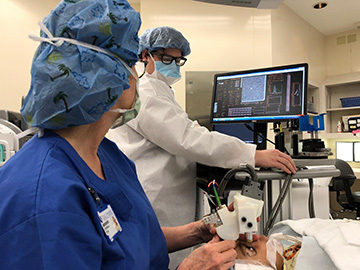
A new hand-held ophthalmology instrument allows imaging of photoreceptors in the human eye. [Image: Theodore DuBose / Duke University]
Sina Farsiu and his colleagues from Duke University, USA, have improved on traditional adaptive-optics scanning-laser ophthalmoscope (AOSLO) design by compacting the technology into a hand-held probe—the HAOSLO—that can image photoreceptors in the human eye (Optica, doi: 10.1364/OPTICA.5.001027).
The key to the streamlined HAOSLO is a new computational algorithm that replaces the bulky AO wavefront sensor and deformable mirror. Custom optics—in the form of a miniaturized deformable mirror and 2-D microelectromechanical systems (MEMS)—further shrink the device and optimize its imaging power. The result, say the researchers, is a hand-held imaging probe that can compensate for subject and user movement, unlike the traditional AOSLO, without sacrificing image quality.
Based on the results of a recent clinical trial, the researchers believe that HAOSLO could someday be used to help clinicians diagnose eye diseases and brain injuries in adults and infants in a variety of clinical and field settings.
Replacing hardware with software
In traditional AOSLO, ophthalmology images are sharpened with a physical device called a wavefront sensor, composed of a large deformable mirror that changes shape in response to wavefront distortions. This instrument is so large that it requires the people being imaged to sit still in an upright position for several minutes, thus making it impractical to use on babies, young children and adults with cognitive or mobility issues.
By creating an algorithm to replace traditional AOSLO’s large wavefront-sensing system, the Duke team was able to dramatically shrink the imaging probe. The new algorithm, unlike previous wavefront-sensor-less AO algorithms, assumes a variable rather than a static optimal deformable-mirror shape. The researchers say that the random nature of the algorithm “allows for dynamic correction, which is necessary to correct for hand motion.” The new algorithm also speeds up imaging without sacrificing resolution.
In addition to the wavefront-sensor-less AO algorithm, the team also improved the optical, signal-processing and mechanical design of the instrument with a 10.5-mm miniaturized deformable mirror and MEMS scanning. The resulting HAOSLO weighs 200 g and measures 10 × 5 × 14 cm.
The portability of HAOSLO could make it useful in an operating room during surgery, or on a sports field to help detect brain trauma in athletes. The researchers also foresee HAOSLO as a tool to better image the eyes of babies, particularly for premature newborns because they have a greater risk of developing eye diseases that could lead to blindness if not diagnosed and treated quickly.
Testing HAOSLO in babies and adults
The Duke team tested HAOSLO in a clinical trial with 12 adults and 2 children. The team showed that HAOSLO could produce high-resolution images of the cones (a type of photoreceptor cell) closest to the fovea (an area at the back of the retina with densely packed cones). And, to the best of their knowledge, the researchers also produced the first AO-enhanced images of cones in infants.
They were also able to image cone density at an eccentricity to the fovea of 1.4 degrees. (The eccentricity value of a circle is zero. Therefore, a value close to zero means less distortion because the image “circle” isn’t stretched or squeezed.) Previous AOSLO designs could image cone density at 3.9 degrees eccentric to the fovea.
The team has made HAOSLO mechanical designs, computational algorithms and control software open-source so that other scientists can adapt the new system for a variety of applications in ophthalmic imaging: http://people.duke.edu/~sf59/HAOSLO.htm.