
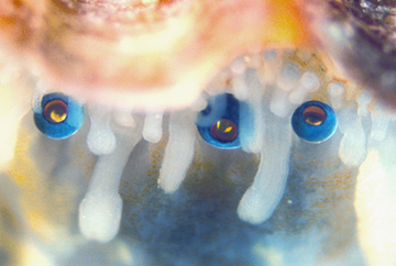
The eyes of the scallop Pecten maximus are nestled amid the tentacles in the mantle tissue that lines the organism’s shell. [Image: Dan-Eric Nilsson, Lund University]
A research team from Israel and Sweden has taken a deep dive into the unusual visual world of the scallop—with some surprising results (Science, doi: 10.1126/science.aam9506). Using a variety of imaging techniques, including cryo-electron microscopy (whose originators received the 2017 Nobel Prize in Chemistry), the scientists have revealed that the visual system of these marine bivalves, which can sport up to 200 individual eyes, relies on a hierarchical array of micrometer-scale multilayer mirrors that, according to the researchers, bear “a striking resemblance to the segmented mirrors of reflecting telescopes.” The team believes that insights from its work on scallops could spur novel approaches to bio-inspired optics for imaging and sensing.
Mirrors, not lenses
It’s been known for several centuries that the Pecten scallop, along with several other marine invertebrates, has a visual system that uses mirrors rather than lenses to focus the picture. In the 1960s, the British neuroscientist Michael Land sussed out the basics of how the system works: Each of the organism’s nearly 200 eyes, which are roughly 1 mm in diameter, includes a tiny concave mirror at the rear of the eye that focuses and reflects light onto a retina positioned above the mirror.
Land speculated that the mirrors were made of a crystalline form of guanine (also one of the four nucleotide bases in DNA). But he was unable to confirm the composition and structure of the mirrors, as their delicate structure could not stand up to the sample prep required for transmission electron microscopy, the only method available at the time.
A world of complexity
The team in the current work, including scientists from the Weizmann Institute of Science, Rehovot, Israel, and Lund University, Sweden, overcame that obstacle by leveraging cryo-scanning electron microscopy (cryo-SEM). In that technique, the tissue to be investigated is flash-frozen, and can thus hang onto structures that would be lost in the dewatering sample preparation required for conventional electron microscopy.
The cryo-SEM opened up the world of complexity that evolution has placed at the back of each of the scallop’s many tiny eyes. Far from being a simple mirror, the reflector at the back of the eye consists of a tiled, segmented array of hundreds of micrometer-scale, multilayer reflecting guanine crystals, analogous to the segmented arrays used by modern giant reflecting telescopes to capture light from distant stars.
Sweating the (optical) details
Equally remarkable is how evolution has managed the optical details. The guanine forming the tiny mirrors (known as β-guanine) grows in a square crystal morphology that differs from the more conventional prismatic crystal form; this allows it to present the crystal face with the highest refractive index on the mirror face, “creating a highly reflective surface,” according to the scientists. The square morphology also allows the crystals to tile efficiently in “an almost perfectly tessellated mosic” that minimizes edge defects that would cause optical aberrations and losses.
The research team used the data on the layered crystals and their configuration to create a simulated reflectance spectrum, which showed a pronounced peak in the blue-green region of around 500 nm. That’s dead on target with the peak in the irradiance spectrum for the light that reaches down into the scallop’s habitat and that is most efficiently processed by the organism’s retina. And the team used X-ray micro-computed tomography to determine the 3-D shape of the mirrors in detail, followed by ray-tracing simulations that showed that the focal point of the mirrors changes to different parts of the retina with changes in the angle of incidence. That allows the scallop’s brain to manage input from both the central and peripheral vision fields of the organism.
Toward new bio-inspired optical elements
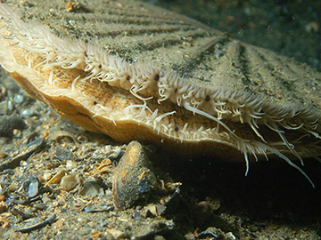
The scallop Pecten maximus. The organism’s eyes are the dark spots amid the tentacles lining the shell opening. [Image: Ceri Jones, Haven Diving Services]
So why does the scallop have up to 200 of these complex eyes, arrayed amid the tentacles that line mantle tissue along the organism’s shell opening? The researchers suggest that the bivalve’s neural processing equipment may integrate the input of the widely dispersed individual eyes (which send back pictures of varying quality, depending on the angle of incidence), to make the overall picture much sharper and better in depth of field than what could be accomplished by any single eye.
It’s a visual approach remarkably different from the one humans have inherited—and, the scientists believe, it could have practical value. Understanding the scallop’s complex visual strategies, the team concludes in the study, “provides inspiration for the development of compact, wide-field imaging devices derived from this unusual form of biological optics.”