
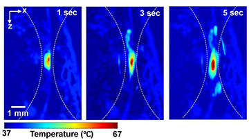
Photoacoustic imaging shows the absolute temperatures elevated by high-intensity focused ultrasound in small animal models at (from left) 1, 3 and 5 s. [Image: Junjie Yao, Duke University]
Measuring overall body temperature is one way to assess a person’s or animal’s general health. However, when administering focused thermal treatments to combat tumors, medical professionals need to know the temperature of the small regions of targeted cells compared with the rest of the surrounding tissue.
Photoacoustic imaging—in which non-ionizing laser pulses heat biological cells, converting some energy into ultrasound waves—can distinguish between relative temperatures of nearby tissues, but alone cannot provide an absolute measurement deep inside an animal’s body. Calibration attempts are difficult to carry out, especially while heat treatment is killing malignant cells or otherwise affecting nearby tissue.
Now, researchers based at a U.S. university have tweaked the basic photoacoustic imaging method to make it a technique for measuring absolute temperatures of small tissue regions (Optica, doi:10.1364/OPTICA.6.000198).
TEMPT
Junjie Yao and his biomedical engineering group at Duke University named their method thermal-energy-memory-based photoacoustic thermometry, or TEMPT, because it builds up information from a sequence of measurements over time. Instead of a single laser pulse, the team aims a burst of nanosecond laser pulses at the target cells. The pulses hit the tissue within the window of its thermal relaxation time, which is typically less than 1 s long.
For each laser pulse, the researchers measure the photoacoustic response of the target tissue. The first measurement records the baseline temperature of the cells; the subsequent ones analyze the cumulative heating of the tissue from each laser pulse. Mathematical modeling provides the estimates of absolute temperature—by taking ratios of the photoacoustic measurements, many of the otherwise unknown or irrelevant quantities, such as the number of photons delivered with each pulse, simply cancel each other out.
Applications in oncology
According to the researchers, TEMPT can be used to map the temperatures of cells during thermal ablation of cancer tissue—heating up the malignant cells with ultrasound or radio waves until the cells die. Obviously, a physician aims to ablate only the cancerous cells, not the healthy tissue, so it’s essential that the temperature stays in the most efficient range.
The group first tested TEMPT on meat slabs before attempting temperature measurements while carrying out the procedure known as high-intensity focused ultrasound (HIFU) on the hind leg of a living mouse. The researchers mapped out the increase in temperature around the target spot over a 5-s period.
Two of the team members are also affiliated with Tsinghua University in Beijing, China.