
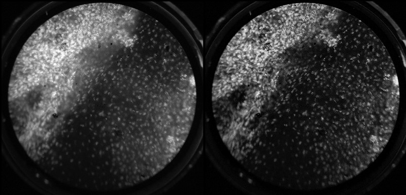
Nuclei from cervical tissue (white spots) imaged using Rice University’s differential structured illumination microendoscopy (DSIMe). DSIMe removes out-of-focus light to improve image resolution (right) when compared with a wide-field HRME image without DSIMe (left). [Image:Pelham Keahey/Rice University]
Researchers from Rice University, Houston, Texas, USA, have developed and demonstrated a distally mounted rotating glass grating that, coupled with an existing endoscope, may boost capabilities for imaging cancers deep within the body (Proc. Natl. Acad. Sci. USA, doi: 10.1073/pnas.1613497113).
Led by OSA Fellow and 2016 MacArthur Fellow Rebecca Richards-Kortum, the team says its new imaging method removes the out-of-focus light scattered by soft tissue. The device could, according to the scientists, be used to improve pre-surgical detection of tumor margins—that is, the border between normal and cancerous cells—to improve the chances of the complete surgical removal of the cancer.
Removing out-of-focus light
Richard-Kortum’s high-resolution microendoscope (HRME) has been used to image—in real time—individual nuclei in esophageal-cancer cells. (Irregularly spaced, large nuclei are a sign of abnormal and possibly cancerous cells.) However, for tissues deeper in the body, the fluorescent dyes used to stain the cells’ nuclei throw back “so much out-of-focus light that it’s difficult to get a clear image,” says Pelham Keahey, first author and a member of Richards-Kortum’s team.
To solve this out-of-focus light problem, Richards-Kortum and Keahey, along with Preetha Ramalingam and Kathleen Schmeler, needed to find a way to overcome the exposure times required for confocal imaging, as well as the multiple images needed for structured illumination—optical techniques that can remove scattered light but are not ideal for non-invasive imaging in live, moving patients. With this in mind, the team needed a solution that would not add bulk to the thin front end of the fiber optic cable inserted into the patient.
The solution they came up with is a rather backward version of structured illumination called differential structured illumination microendoscopy (DSIMe). With DSIMe, the team added the optomechanical devices for eliminating out-of-focus light to the back of the fiber optic cable, instead of adding bulk to the front end. The star of DSIMe is a spinning, patterned-glass grating. The researchers say the modifications do not add bulk or sacrifice the imaging speed of the portable HRME.
DSIMe in action
The spinning, reflective-glass grating interacts with light from a high-powered LED. The modulated illumination pathway is then imaged onto the fiber optic cable that is exposed to the tissue to be imaged. The modulated light from the cable creates a striped pattern on the tissue stained with a molecular contrast agent. The return signal from the dyed nuclei returns along the same optical path as the outgoing light and is imaged onto the grating.
Light from in- and out-of-focus optical planes pass through the grating and are imaged onto a rear-mounted charge-coupled device (CCD), creating a wide-field image. Another image is formed by light from optical planes, past the point where the grating image on the optical fiber has decayed. This light is reflected off of the spinning grating and onto a front-mounted CCD. A real-time image-stabilization algorithm takes the two CCD images and removes the out-of-focus light to create an image with better contrast.
These sharper images can be displayed on a computer monitor or on smart glasses. The smart-glasses interface, the researchers say, can be used “to relay images directly to clinicians during in vivo operation of the device.”
The team demonstrated DSIMe with fluorescent microspheres on optical slides; imaging of mouse stomachs and colons; and, most recently, in a clinical pilot study on patients undergoing surgery due to cervical cancer. Richards-Kortum and her team say that the images acquired from the clinical pilot study using DSIMe “show greater contrast than standard microendoscopy, improving the ability to detect cellular atypia associated with neoplasia.