
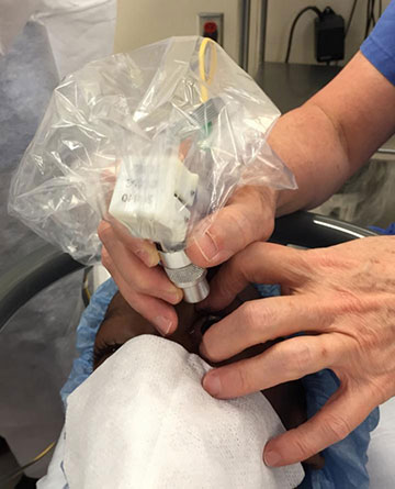
The new handheld probe in use on a human test subject. [Image: Joseph Izatt, Duke University]
Optical coherence tomography (OCT), adaptive optics and other technologies have opened up a world of possibilities for imaging the retina, right down to the photoreceptor cells (rods and cones) that are the gateway to vision. But the equipment for cellular-resolution imaging is bulky, heavy and inconvenient, making it all but useless for addressing one key problem: tracking how the retina grows and matures during infancy and early childhood.
Now, a research team led by OSA Fellow Joseph Izatt of Duke University, along with Duke colleagues Cynthia Toth and Sina Farsiu, has leveraged several different innovations to build a new, lightweight handheld imager that can grab retinal images in young patients, at cellular resolution (Nat. Photon., doi: 10.1038/nphoton.2016.141). The new device—characterized in a press release as “about the size of a pack of cigarettes,” and weighing “no more than a few slices of bread”—could allow clinicians and researchers, for the first time, to bring the retinal imaging advances of recent years to bear in understanding the rapid changes the retina undergoes during infancy and childhood. It could also shed light on how retinal diseases develop during these crucial early years.
Bulky current systems
Current diagnostic tools for retinal imaging have allowed great strides in understanding retinal photoreceptor structure in adults, who will sit still in fixed positions long enough to collect the data (which, with some systems, can take several minutes). But infants and young children aren’t so cooperative.
Handheld retinal imaging systems do exist, but they have lacked the resolution to suss things out at a cellular level. And while several teams (including Izatt’s) have developed compact and even handheld OCT and scanning laser ophthalmology (SLO) systems capable of visualizing cone photoreceptors, these have still proved too heavy and bulky to hold in place for use on children.
Addressing the pain points
To address these problems, the Duke team—which also included recent Ph.D.s Francesco LaRocca and Derek Nankivil, and current grad student Theodore DuBose—focused on three key elements that have added bulk to existing systems. For starters, they created a new design for the relay telescope that images the scanning plane into the patient’s pupil. The new design, which uses converging rather than collimated light, allowed up to a 50 percent reduction in length relative to the standard 4f imaging telescopes used in previous designs.
The team also replaced the two large galvanometer-based optical scanners with a single high-speed, 2-D micro-electromechanical (MEMS) scanner. And first author LaRocca designed custom lenses for the instrument, including an asymmetric triplet lens that minimizes induced monochromatic aberrations and corrects for chromatic aberrations across the spectrum of illumination.
Opening up new research opportunities
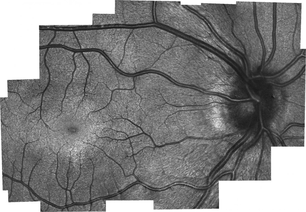
An image of the retina, taken with the handheld probe, provide a clear enough view of the cone photoreceptors (white dots) to allow the cone density to be calculated. [Image: Joseph Izatt, Duke University] [Enlarge image]
The result is a combined SLO/OCT probe that can fit comfortably in the hand, weighs in at a mere 94 g, and can conceivably be held in place long enough to gather data even from squirming young patients. Initial tests (done on children who were undergoing other eye exams under anesthesia) captured sharp images in which cones were plainly visible, allowing for calculations of cone density relative to normal adult eyes. And, according to lead author La Rocca, the tests “showed different microscopic pathological structures that are not normally possible to see with current, lower-resolution clinical-grade handheld systems.”
The group is now working with clinicians at the Duke University health service to get feedback that can be for an improved, next-gen device. The researchers expect the probe to “have a significant impact on our understanding of retinal development, maldevelopment and early onset of diseases during human growth.”