
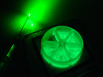
Bioluminescence from cells cultured in a multi-electrode array, next to a fiber optic cable. Credit: Jack Tung
Researchers from Emory University and the Georgia Institute of Technology (USA) report a new method for optogenetic inhibition deep within the brain—untethered by fiber optic filaments. Instead of using fiber optics to deliver light into the brain, the scientists use a glowing protein as an internal light source that can be turned on by a chemical injection (Sci. Rep., doi: 10.1038/srep14366). When activated by the light, these “inhibitory luminopsins” inhibit neuron activity. According to the scientists, this non-invasive technique expands the optogenentic toolbox and could make optogenetics research in freely moving animals more practical.
In most optogenetics experiments, light is delivered via fiber optics to neurons that have been genetically engineered to release light-sensitive proteins called opsins. The opsins either activate or inhibit the neurons from which they are expressed. Although they are widely used, surgically implanted fiber optic filaments aren’t ideal for deep-brain studies because their bulk limits their reach in brain tissue, can impede animal movement, and may also introduce infection.
To create their new optogenetics tool, Robert Gross and his colleagues at Georgia Tech and Emory took luciferase (an enzyme that glows when combined with luciferin) and coupled it to an inhibitory opsin. They demonstrated their hardware-free inhibitory luminopsins in vitro and in vivo. For the in vitro study, the researchers showed that their inhibitory luminopsins suppressed neuron activity in cultured cells when exposed to both luciferin and an external fiber-optic light source. One of their in vivo studies demonstrated the inhibitory luminopsin’s ability to switch off half of the globus pallidus–a section of the brain that controls motor function—in awake and freely moving rats. After the rats were injected with luciferin, they rotated in only one direction, a sign that half of the globus pallidus section was “switched off.” This is the same motor behavior that occurs when half of the globus pallidus is damaged. They also demonstrated that the inhibitory luminopsin could suppress hippocampus activity in live, but anesthetized rats.
The authors say their new class of hardware-independent optogenetic probes “also permits multi-modal (i.e., optical and chemical) methods of neuromodulation that can result in varying temporal effects.” Gross’s team is now using the inhibitory luminopsins to hopefully find ways to stop or prevent generalized or seizures and seizures that involve several areas of the brain.