
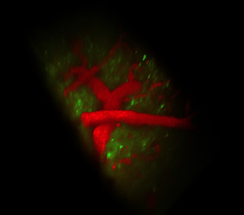
SCAPE imaging of the living brain of an awake mouse at 10 volumes per second. Green GCaMP marks apical dendrites of layer 5 neurons, and red shows Texas red dextran in the vasculature. Credits: Elizabeth Hillman and Clay Lacefield
Biomedical engineers at Columbia University Medical Center (CUMC, USA) have reported a three-dimensional microscopy technique called swept, confocally-aligned planar excitation (SCAPE) microscopy, which allows imaging of living volumetric samples at ultrahigh speed. Elizabeth Hillman, associate professor of biomedical engineering at Columbia Engineering and radiology at CUMC, developed a novel approach that improves upon other techniques such as laser scanning confocal, two-photon and light-sheet microscopy to deliver 10 to 100 times faster 3-D imaging speeds of living samples.
Conventional light sheet microscopy commonly uses two-objective, orthogonal illumination, and either physical translation of the sample or complex synchronization of the illumination and detection planes. Together, these constraints limit both the imaging speed and the type of sample that can be imaged. SCAPE microscopy, by contrast, uses an angled sheet of swept light, a single-objective lens, and confocal descanning to obtain completely translationless 3-D imaging at a rate exceeding 20 volumes per second.
Hillman and colleagues successfully imaged, for the first time, spontaneous neuronal firing in the rodent brain, single cells in the beating heart of zebrafish and crawling fruit-fly larvae (Nat. Phot., doi: 10.1038/nphoton.2014.323). The combination of single-objective lens and swept light sheet makes SCAPE fast, easy to use and inexpensive. “We think SCAPE will be transformative in bringing the ability to capture high-speed 3-D cellular activity to a wide range of living samples,” said Hillman.
Hillman hopes the advance may someday help scientists visualize and monitor the secrets of live brain activity, which was barely imaginable a few years ago. The new technology has been made available for licensing from Columbia Technology Ventures.