
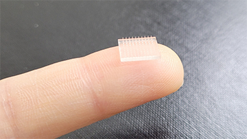
Scientists at the University of North Carolina at Chapel Hill and Stanford University used 3D printer to produce microneedle vaccine patch that dissolves into the skin to boost immunity. [Image: University of North Carolina at Chapel Hill]
While vaccines are traditionally given as injections into the muscle, delivery through the intradermal route—in other words, between the superficial layers of the skin—has several notable advantages. Human skin is rich in immune cells, which means a much smaller dose can stimulate an equal immune response, reducing overall costs and improving access to vaccines in places with limited supply.
Now, researchers from Stanford University, USA, and the University of North Carolina at Chapel Hill, USA, have used a fast, light-based 3D-printing process to create an intradermal microneedle vaccine patch (PNAS, doi: 10.1073/pnas.2102595118). When tested on mice, the patch resulted in more potent immune responses when compared to traditional vaccination routes.
Faster 3D printing with light
Previously, Joseph M. DeSimone and his colleagues invented a novel approach to 3D printing called continuous liquid interface production (CLIP) that boasts rapid manufacturing speeds and high feature resolution. In 2016, they employed CLIP to successfully print microneedle patches with tunable geometries in the span of minutes.
CLIP works by projecting a continuous sequence of UV cross-sectional images—generated by a 385-nm light-emitting diode (LED) and digital light processing imaging unit—through an oxygen-permeable, UV-transparent window. The window is located at the bottom of a liquid resin bath, so that the cross-sectional images are able to cure the resin and build up the object on a steadily rising support plate.
“We can grow these objects continuously, with no sticking to the window. The resin renewal happens at the gap between the building part and the window created by the oxygen coming through,” said DeSimone, study author and professor of translational medicine and chemical engineering at Stanford University. “We call that [permanently liquid] gap the ‘dead zone.’ So it’s a breakthrough in how 3D printing is done.”
A boost in immune response
The researchers had to significantly refine their CLIP printer to achieve the proper length scale for vaccine delivery via microneedle patch, which consists of an array of micrometer-sized solid needle projections. But the principles behind CLIP remained the same, and the dead zone allowed for the creation of much smaller and more delicate structures. A traditional layer-by-layer stereolithography approach, on the other hand, would require breaking off of structures from the window each time.
DeSimone and his colleagues printed a 10×10 array of faceted microneedles with ridges to increase the surface area and vaccine cargo compared to a square pyramidal shape. When coated with vaccine components for in vivo experiments, the intradermal patch generated an immune antibody response that was 10 times greater than injection into muscle and 50 times greater than a subcutaneous injection in mice.
“This is a whole new day in making microneedles because now you can make pixel sizes even smaller. In the paper, we used a printer with 20-micron-sized pixels. Here at Stanford, we’ve built a new printer with 1.5 micron-sized pixels,” he said. “So we’re making some amazing new generations of microneedles where you can now make all kinds of architected structures and really open up a wide range of performance.”