
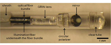
Researchers have developed a small-diameter intravascular catheter that incorporates a fiber bundle, polarizer and GRIN lens to image the reflected speckle patterns onto a CMOS sensor. [Image: Seemantini Nadkarni, Wellman Center for Photomedicine]
Doctors employ a variety of optical tools for cardiovascular imaging to diagnose and treat heart disease, including atherosclerotic plaques, which cause most fatal heart attacks.
Working toward a goal of better predicting and preventing heart attacks caused by ruptured coronary plaques, researchers at Massachusetts General Hospital and Harvard Medical School, USA, now report proof-of-concept, in vivo use of intravascular laser speckle imaging (ILSI) to measure the viscoelastic properties of these lesions (Biomed. Opt. Exp. doi: 10.1364/BOE.418939).
Pinpointing fatal mechanical stress
Measuring the viscoelastic properties of coronary plaques, write the authors, who constitute a team at Harvard/Mass. General’s Wellman Center for Photomedicine, is the “next major hurdle for ILSI in the clinical translational journey.” They write that their results show that this imaging modality will be “crucial in identifying mechanically unstable plaques with a propensity for rupture and subsequent MI [myocardial infarction].”
In an accompanying news release, the authors note that there is compelling evidence that, in addition to microstructural and compositional factors—as measured by other imaging techniques—plaque rupture is also mediated by mechanical factors, particularly peak mechanical stress. This factor is in turn influenced by the viscoelastic properties of the plaque, they write, and thus presented a good target for investigation over the course of several studies.
Measuring viscoelasticity with light
“Reducing mortality from heart attacks in the general population requires a comprehensive screening strategy to identify at-risk patients and detect high-risk vulnerable plaques while they can be treated,” said Seemantini K. Nadkarni, an associate professor of medicine at Harvard, in a press release. “By providing the unique capability to measure mechanical stability—a critical metric in detecting unstable plaques—ILSI is poised to provide a new approach for coronary assessment.”
ILSI is an optical technique capable of quantifying an index of viscoelasticity of the coronary vasculature, the researchers write. Laser speckle refers to a granular pattern formed by the interference of coherent laser light scattered from tissue. Hence, “Speckle fluctuations arise from and are modulated by the Brownian motion of endogenous light scatterers and are in turn closely related to the viscoelastic properties of the tissue microenvironment,” they note in their paper.
The team’s prior experiments with ILSI were conducted ex vivo on diseased aortas from human cadavers and so-called test phantoms. The researchers next developed a flexible, miniaturized optical catheter and tested it on rabbits.
The team describes the ILSI catheter as integrating a small-diameter fiber bundle, a polarizer and a GRIN lens for lighting up the coronary wall and transmitting the laser speckle pattern to an external, high-speed CMOS camera. The instrument was placed in a specially built catheter sheath designed to keep blood from the field of view during imaging.
Testing ILSI under the forces of life
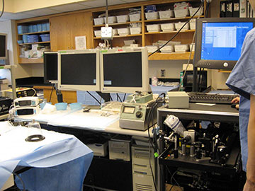
Pictured is the animal catheterization laboratory with the portable ILSI system on the cart (right). [Image: Seemantini Nadkarni, Wellman Center for Photomedicine]
For the current study, the Wellman team grafted human aortic tissue with coronary plaques to vasculature in anesthetized pigs and diverted blood flow through the human aorta. With the ILSI system, the researchers report taking measurements of the coronary plaques under real-life conditions of a breathing animal with a beating heart—motions that may contribute to mechanical stress on coronary lesions leading to rupture.
The procedure setup included an imaging console on a portable cart that housed a 30-mW helium–neon laser source. The team used a high-speed triggerable CMOS camera to record laser speckle patterns at reported acquisition rates of 1,000 frames per second. Meanwhile, other external instruments recorded the swine EKG and/or blood pressure signals during laser-speckle pattern acquisition.
Study results, the team writes, suggest that ILSI could become a viable method for rapid screening of coronary tissue. “We anticipate in the future that ILSI may be applied in clinical interventional cardiology practice for identifying high-risk plaques and guiding plaque stabilization therapies, either as a standalone approach or as a multimodal outfit, combined with other complementary intravascular imaging techniques,” they write.