
![]()
Researchers developed a multimode fiber holographic endoscope for far-field imaging. [Image: Courtesy of Tomáš Čižmár]
Holographic endoscopes consisting of hair-thin multimode optical fibers have emerged as promising candidates for minimally invasive biomedical imaging. Their potential beyond the capabilities of conventional endoscopes, researchers say, has been demonstrated particularly in in vivo animal studies that have captured living neurons and processes of neuronal circuits implicated in neurodegenerative conditions like Alzheimer’s disease.
Now, with the aid of a sweet pepper and a mechanical clock, a research team at Germany’s Leibniz Institute of Photonic Technology reports a proof-of-concept demonstration that would extend the capabilities of commercially available holographic endoscopes to include imaging of a target from a distance, or far-field imaging (APL Photon. doi: 10.1063/5.0038367). The team says its studies have shown that far-field imaging could reduce the endoscope’s already low footprint below 0.1 mm while reaching microscopic resolution at unprecedented depth of tissue.
Imaging from a distance
Coupled with the endoscope’s ability to switch, at the push of a button, between far-field and microscopic imaging modes, the researchers say, distance imaging could significantly broaden use of these instruments for both disease research in animal models as well as potential clinical/diagnostic applications.
The Leibniz team—Ivo T. Leite, Sergey Turtaev, Dirk E. Boonzajer Flaes, and Tomáš Čižmár—focused on imaging targets at macroscopic distances of 200 mm to 400 mm from the distal end of the instrument, and on where they could address limitations such as power efficiency and wavefront control fidelity.
The study shows that you can image objects very far away from the fibers, explains co-author Čižmár, who is also the head of the complex photonics lab at the Czech Academy of Sciences, Czech Republic. “Since you can switch from one working distance to another by a push of a button, the far-field capability will be very useful, for example, to navigate towards suspicious tissue through natural orifices like the gastroenterological tract or lungs before turning your device into a high-resolution microscope working in the proximity of the fiber facet,” he adds.
Holographic wavefront imaging
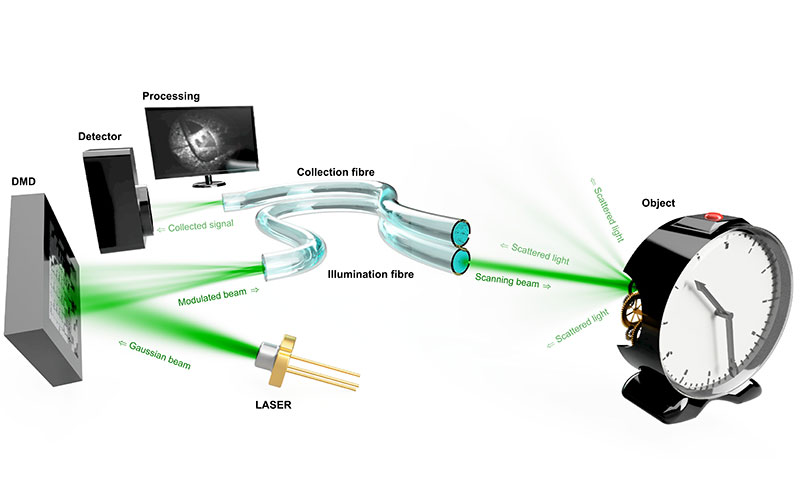
A sequence of holograms displayed by a digital micromirror device spatially shapes the wavefronts coupled into a multimode optical fiber in such a way that a far-field focus scans the distal field of view. [Image: Tomáš Čižmár][Enlarge image]
In holographic wavefront imaging, Čižmár explains, one fiber is used to illuminate the target and the other to collect the backscattered or returning photons. Together, the fibers form a lensless imaging instrument capable of conveying much higher densities of information than conventional endoscopy. But for the technology to be clinically useful, he says, it is important to understand how limitations such as fewer returning photons to the receiving fiber shaft can affect image quality as well as whether the instrument can meet the imaging demands of dynamic environments.
And that is where the pepper and clock come in.
“The versatility of the instrument was demonstrated by imaging complex three-dimensional scenes, particularly the interior of a sweet pepper serving as a phantom for biomedically relevant environments, as well as a functioning clockwork mechanism as an example of an object with dynamic complexity,” the team writes.
Meeting high imaging standards
What’s more, the team reports transmitting 0.1-megapixel images, which reaches the standard of modern video endoscopes. Their work also revealed that field of view, resolution and minimum imaging distance of the endoscope depend solely on the numerical aperture and core size of the illumination fiber.
The most promising use of holographic endoscopes in human medicine is probably in the diagnostics of hard-to-access locations of the body like the ovaries or pancreas, Čižmár says. “When diagnosing suspicious tissues, chemical staining of post-mortem tissues is used. There is a great potential to replace such procedures with purely light-based approaches and implement them in situ.”
But, he emphasizes, applications in animal models are also important. “Should holographic endoscopy assist in animal studies, for example, to uncover secrets behind dementia and enabling development of suitable medications, this by itself would be a huge endorsement of this technology—well worth the investments and efforts.”
Challenges ahead
Challenges ahead for further development of the holographic endoscope technology, Čižmár says, include, for neuroscience, the ability to study fully awake animal models.
“This will enable us to link our observations of what happens in the animal’s brain at the microscopic level with what the animal goes through—orientation in space, emotions, reactions to visual or motoric stimuli and so on—and particularly how all these things change during onset and progression of a neuronal disease,” he says.
But allowing study animals to move around is itself challenging, Čižmár says, because the fiber can get bent, looped or twisted. “This changes the light transport through and impedes the quality of imaging you can get,” he says, adding that the team is working diligently to find a practical solution.