
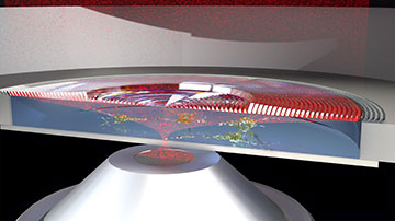
In this cross-section view of a SRS microscope, a red laser beam is strongly focused by a high-NA objective to interact with live cells. The 3D-printed, catadioptric Fresnel lens on top collimates the beam, redirecting it upward toward a photodetector. [Image: © 2020 KAUST; Andrea Bertoncini]
Label-free, fast chemical imaging available with stimulated Raman scattering (SRS) microscopy has found powerful applications in biology and medicine. But a phenomenon known as cross-phase modulation (XPM) can create background signals that ruin the picture.
Now, a 3D-printed lens—reminiscent of the Fresnel lens used in lighthouses—could be used to eliminate XPM and expand the capabilities of this important spectroscopic method, reports a research team in Saudi Arabia (J. Biophoton., doi: 10.1002/jbio.202000219).
Collecting the right vibes
In SRS microscopy, two laser pulses collect molecular vibrational signals from biological samples. The vibrational signals allow SRS microscopes to map the 3D distribution of chemical bonds within a sample with sufficient speed and sensitivity to allow near-real-time imaging, explains co-author Carlo Liberale, assistant professor of bioscience at King Abdullah University of Science and Technology (KAUST) in Thuwal, Saudi Arabia.
The XPM effect occurs, he says, when the two intense laser beams used in an SRS microscope setup interact to create a non-resonant signal indistinguishable from the desired Raman signal from the sample. It can be suppressed using high-numerical-aperture (high-NA) microscope objectives to collect the laser beams after their interaction with the sample, but this implies very short working distances.
To the lighthouse
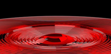
The thin, affordable lens is 3D printed and has the capacity to put live cells under the microscope. [Image: © 2020 KAUST; Andrea Bertoncini]
This limitation became a stumbling block for the researchers. When they started doing live-cell SRS microscopy with an inverted microscope and a stage-top incubator to keep the right conditions for the cells, the incubator was not compatible with the high-NA collection optics.
To reject the XPM background in SRS microscopy, Liberale says, researchers need a way to collect a highly-focused beam over the largest possible angle—that is, the largest NA—but without any special need for imaging quality of the collection optics.
“We just needed a non-imaging optical element,” Liberale says, “which relaxes the requirements for its optical design.”
Enter the lighthouse lens idea. “In its ‘complete’ version, such lenses include both refractive [lens-like] and reflective elements,” Liberale says. “The lens is a catadioptric element. Indeed, lighthouse Fresnel lenses used a catadioptric design to redirect the light of a torch towards distant ships, to signal the presence of land.”
“It’s a very efficient way to collect and redirect light from a source that emits rays over a very wide angle—in this case, the torch,” says co-author Andrea Bertoncini, a member of Liberale’s group. “In our case, this wide angle is with the laser beam to be collected, which emerges highly divergent after having been tightly focused on the sample.”
Making it all fit
The next challenge for the KAUST team was fitting the 3D-printed microlens to the microscope setup.
“Initially, we struggled to find a mounting strategy that would guarantee a precise working distance, and that could fit the lens inside the incubator,” Liberale says. “The solution was hiding in plain sight: We redesigned the lens to have a working distance matching the depth of the petri dish well.”
This solution of how to incorporate the lens, he says, stems from the fact that, “beyond developing tools, we directly work with applications—in this case live-cell microscopy—and so we have ended up with an arrangement which ‘reuses’ the petri dish also as part of the optical configuration.”
A two-photon technique
The team’s lens was printed with a technology based on two-photon lithography, first presented about 20 years ago. According to Liberale, the technique allows 3D printing with a spatial resolution better than popular consumer or professional 3D printers. Recent advances have provided new printable materials and printing configurations suited to fabricating micro-optics, he adds. To further enhance the collection capability of the team’s lens, Bertoncini says, an antireflective coating could be added to the surface of its 3D micro-elements.
“Our lens is thin, reusable and easily integrated just by laying it on top of the sample,” Bertoncini says, and it could be incorporated into commercial laboratory equipment for SRS microscopy or other applications.
Unfolding more applications
“Imaging techniques employing highly-focused and thus highly divergent beams could indeed benefit from the high-NA collection capability of our lens,” Liberale says. “We think it could find applications not only in bioimaging but also in material science, for example, the collection of photons from quantum emitters.”
For their study, the researchers imaged liver cancer cells, but Liberale says SRS microscopy is a powerful bioimaging tool for a variety of biomedical studies. “We expect the range of applications to expand,” says Liberale. “We believe that the improvement in SRS microscopy provided by our lens could benefit all experiments for which it’s crucial to image live-cells under precise environmental conditions.”