
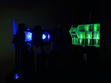
Researchers developed a new multifocus technique that uses a z-splitter prism (right) to split detected light in a standard microscope. [Image: Sheng Xiao, Boston University]
Optical microscopy is an essential tool for producing sharp, in-focus 2D biological images, but it can be tricky to achieve that same sharpness over extended depth ranges with a standard camera-based microscope. Now, researchers at Boston University, USA, have demonstrated a high-speed, large field-of-view (FOV) multifocus microscopy technique based on a z-splitter prism that can be readily applied to a range of biomedical and biological imaging applications (Optica, doi: 10.1364/OPTICA.404678).
The researchers say their method is versatile, flexible and fast, with a basic design that can be assembled using off-the-shelf components and easily added to most camera-based microscopes.
Going deeper
The most common optical-microscopy technique is to use a digital camera to record images at a single focal plane. It’s a simple, affordable, low-noise solution that—thanks to modern sCMOS sensors—offers lightning-fast speeds and high pixel counts. Standard camera-based optical microscopes, however, only provide a sharp image over a very thin 2D plane. Acquiring images at different focal depths usually requires axial scans, which sacrifice either imaging speed or system complexity.
The Boston team wanted to find a simple and fast way to obtain 3D information with standard microscopy. At the crux of the researchers’ multifocus technique, says coauthor Sheng Xiao, is a low-cost, light-efficient z-splitter prism. As implied in its name, a z-splitter prism splits the microscope detected path into multiple paths that are directed onto a single camera with increasing delays. Each path corresponds to a different focal plane and can be simultaneously imaged in one frame.
Compared with traditional multifocus techniques that use beam-splitting optics, says Xiao, the team’s z-splitter-based design is simpler, more achromatic and more versatile, so it can be easily applied to a variety of imaging modalities and can be assembled entirely from off-the-shelf parts. “Our system is also able to provide much larger FOV,” says Xiao, “allowing for imaging hundreds of neurons across volumes for brain function studies, or imaging freely moving organisms in their natural state for animal behavioral studies.”
Shifting focus
Another aspect of the volumetric imaging strategy is a clever deconvolution algorithm, which tackles the common problem of low image contrast caused by out-of-focus backgrounds when imaging thick fluorescent samples. Traditional algorithms, according to Xiao, only account for out-of-focus background contributions in the volume being imaged, failing to remove the far-out-of-focus background outside of that volume.
The researchers’ extended-volume 3D (EV-3D) deconvolution strategy, on the other hand, explicitly extrapolates fluorescent signals beyond the imaging volume, allowing the team to more accurately estimate and remove such far-out-of-focus background, and to ultimately improve the image contrast and signal-to-noise ratio. “This is particularly beneficial in imaging applications involving thick samples,” Xiao says, “where the fluorescence labeling is dense, as is often encountered, for example, when imaging brain tissue.”
In experiments, the team applied its versatile method to fluorescent, phase-contrast and dark-field imaging, capturing large-FOV brain images and monitoring freely moving organisms in 3D and at video rate. The researchers swapped in three different z-splitter configurations to a standard widefield microscope to prioritize either speed or imaging volume, depending on the application.
More modalities
Down the road, Xiao expects that the team’s z-splitter method could be used for brain imaging to help scientists understand brain function and cure neurological diseases, as well as for small-animal behavioral studies.
Currently, Xiao and his colleagues, including lead researcher Jerome Mertz, are working to expand the system to even more imaging modalities and contrast mechanisms. The goal, Xiao says, is to “image an even wider range of samples, making our technique a more general platform for high-speed 3D imaging for biological and biomedical researches.”