
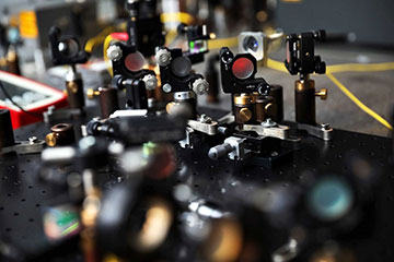
Researchers used a technology borrowed from quantum optics to perform optical coherence tomography (OCT) with much lower light powers than previously possible. [Image: Andrzej Romański]
Strict safety limits on non-ionizing radiation often restrict the use of optical coherence tomography (OCT) in medical diagnostics. By pulling a detection scheme out of the quantum-optics toolbox, researchers in New Zealand and Poland have demonstrated OCT imaging that requires a tiny fraction of the light power of existing systems (Opt. Lett., doi:10.1364/OL.393162).
Safety first
Clinicians often rely on OCT systems, which illuminate subjects with a near-infrared semiconductor light source, to provide high-resolution cross-sectional images of tissue. National and international standards organizations place upper bounds on the emitted power of these devices, especially when used on highly sensitive body parts such as the retina. Sometimes the “photon budget” becomes so tight that users cannot collect enough backscattered light from tissues to produce a clear image.
A group led by physicist Sylwia Kolenderska of the University of Auckland, New Zealand, realized that if the detectors in OCT clinical systems could collect more photons, the resulting images would gain more resolution. The researchers drew from the principle of dispersive Fourier transformation, in which a fiber spool introduces a wavelength-dependent time delay and a single-pixel detector picks up the signal. Scientists already use the technique in various quantum-optics applications and even in some OCT systems that use a supercontinuum light source.
High sensitivity at low intensity
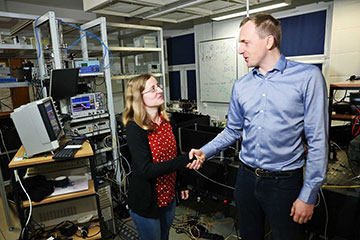
Pictured are researchers Sylwia Kolenderska, University of Auckland, New Zealand, and Piotr Kolenderski, Nicholaus Copernicus University in Toruń, Poland. [Image: Andrzej Romański]
Kolenderska and her colleagues set up an experimental OCT system with a pulsed laser emitting at a central wavelength of 1550 nm. After interacting with the target sample through an interferometric-style setup, the pulses passed through a 5-km-long spool of single-mode fiber. At the spool’s output end, a superconducting single-photon detector, with a peak quantum efficiency of about 65% at wavelengths around 1550 nm, registered and time-stamped each pulse.
The dispersion created by the long light path within the fiber spool delays each wavelength by a different amount of time, so the time measurement by the single-photon detector provided a spectrum of the light entering the fiber spool.
The team imaged two types of targets: a stack of thin sheets of different types of glass and a piece of onion. It took about 15 minutes to acquire each image. More important, each image required only about 10 pW of input light—five orders of magnitude below safety standards.
Benefitting from deep learning
Before the device can be developed for clinical applications, scientists still need to figure out how to remove the parasitic self-interference artefacts that showed up in the images of the onion, which, as biological material, has a more complicated internal structure than parallel sheets of glass. Deep-learning algorithms may help with this.
Kolenderska and one of her co-authors, University of Auckland physicist and OSA member Frédérique Vanholsbeeck, are affiliated with New Zealand’s Dodd-Walls Centre for Photonic and Quantum Technologies. The third team member, Piotr Kolenderski, is at Nicholaus Copernicus University in Toruń, Poland.