

Artist’s rendering of the terahertz field transmitted by an abstract object. Researchers at the University of Sussex, U.K., have demonstrated a setup that allows time-resolved ghost imaging in the terahertz band, opening up prospects for terahertz hyperspectral imaging and detailed chemical and physical analysis of a range of samples. [Image: University of Sussex]
Building on previous theoretical work, a research team at the University of Sussex, U.K., has demonstrated a prototype camera that uses nonlinear “ghost” imaging to perform hyperspectral imaging in the terahertz frequency band (Optica, doi: 10.1364/OPTICA.381035). The researchers believe that the camera, which reportedly can capture terahertz field details at a subwavelength scale, could open up a range of applications in security, bioimaging, manufacturing and more.
The terahertz difference
Terahertz radiation—sometimes referred to as the “far infrared,” and covering the wavelength region from 30 µm to 3 mm—has some distinct potential advantages in imaging and spectroscopy. As non-ionizing radiation, terahertz waves can penetrate many materials without harming them, and can return information on structure and composition even from fragile living materials.
In particular, terahertz time-domain spectroscopy (TDS), which uses sampling with a variable time delay to return complete information on both phase and intensity, has emerged as a powerful scientific tool for getting at the precise internal chemical and physical characteristics of samples. One reason is that many materials have fundamental molecular resonances in the terahertz frequency band.
The detection quandary
Such capabilities could have significant applications in broader commercial areas such as clinical bioimaging, auto sensors and airport security. But the big practical challenge has been on the imaging side. The most cost-effective devices for detecting terahertz fields have tended to be relatively crude thermal cameras, which capture only the time-averaged intensity distribution, without any phase information. And many of the detector implementations that can return full complex-field information—intensity and phase—have tended to be complicated and potentially impractical.
One possible resolution to the quandary lies in so-called ghost imaging, which has seen rapid recent advances in research labs (see “Ghost Imaging,” OPN, October 2016). In the “classical” version of ghost imaging, an object is repeatedly illuminated by different, computationally generated optical patterns, and the transmitted or reflected light is captured by a single-pixel bucket photodetector. A weighted sum of the scattered signal can then be correlated with the spatial distribution of the input structured light, and an image of the object can be computationally reconstructed.
In principle, ghost imaging could be combined with terahertz TDS to allow full field analysis with a very simple detector setup. But here, another practical challenge emerges: the controllable structured-light patterns on which ghost imaging depends are tough to create in the terahertz realm. That’s because the usual tools for generating structured light in the optical domain, such as spatial light modulators (SLMs), are harder to implement in terahertz.
Toward terahertz time-resolved ghost imaging
In a previous theoretical study, published in 2018, the University of Sussex team behind the new work, led by OSA member Marco Peccianti, hit upon a way around this roadblock. Instead of trying to create structured terahertz light for ghost imaging directly, the structured light could be created using an ordinary SLM in the optical domain, and then converted into terahertz structured light using a nonlinear crystal. Because the original structured light is built in the higher-frequency optical domain, the approach opens up the possibility of including detail at the subwavelength scale in the terahertz band.
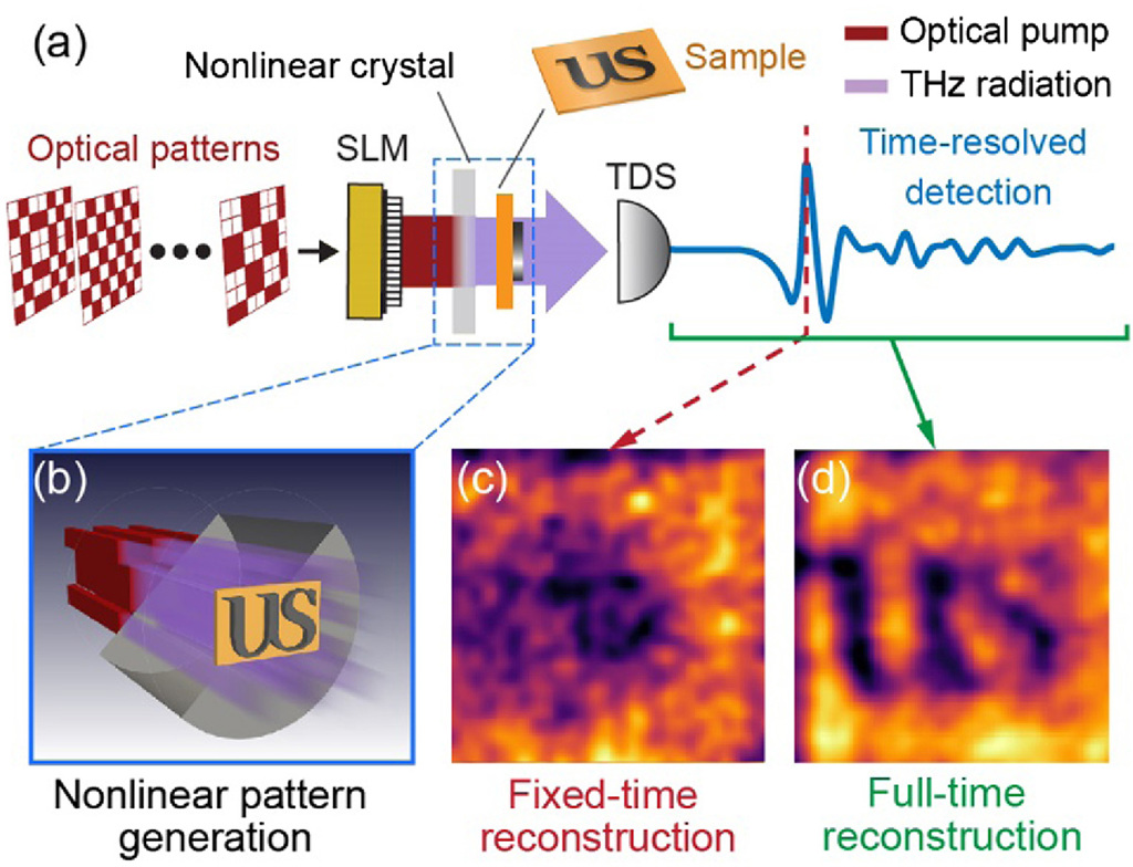
The Sussex team’s setup uses a nonlinear crystal to convert 800-nm patterned light into terahertz patterned light, for use in a time-resolved ghost-imaging setup (a and b). The image of the object, which is not apparent in a fixed-time reconstruction (c), becomes fully visible in the team’s time-resolved detection scheme (d). [Image: L. Olivieri et al., Optica, doi: 10.1364/OPTICA.381035] [Enlarge image]
The team’s modeling in the 2018 study suggested that this approach, which the researchers dubbed time-resolved nonlinear ghost imaging (TNGI), could reconstruct the full optical response of the sample at terahertz frequencies. That, they argued, could enable “a complete hyperspectral imaging approach” that would allow deep investigation of complex, inhomogeneous targets using off-the-shelf optical components.
Leveraging nonlinear optics
In the new study in Optica, the team has now implemented this theoretical design. Their setup begins with a spatial light modulator, which impresses a series of computationally generated intensity patterns on an 800-nm, pulsed optical beam, with a repetition rate of 1,000 pulses per second and a pulse duration of 75 fs. A nonlinear ZnTe crystal next converts that structured light into a structured terahertz field distribution, which interacts with the sample. The scattered field from the sample is then captured using a bucket photodetector that’s been set up to do time-domain spectroscopy analysis, and the full field is reconstructed computationally using ghost-imaging techniques.
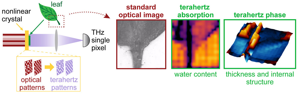
The Sussex team used the setup to perform terahertz hyperspectral imaging of a leaf specimen. The time-resolved approach allowed the recovery of phase information that enabled imaging of the leaf's internal structure. [Image: University of Sussex] [Enlarge image]
Using the setup, the team was able to perform hyperspectral imaging of simple metallic masks, using its time-resolved approach to pull out complete field information and capture subwavelength features of the objects being imaged that were invisible when using fixed-time approaches. The researchers were also able to capture hyperspectral images of a biological sample (a leaf, in various stages of dessication).
In a press release accompanying the research, team leader Marco Peccianti noted that, through its ability to preserve complete field information, terahertz TNGI allows “the fingerprint of all the details of the image” to be pulled out in a way that enables deep investigation of the sample. The next phase of the research, according to one of Peccianti’s coauthors, OSA member Juan S. Totero Gongora, will focus on refining and speeding up the system, to take it closer to “to real-world applications like airport security, intelligent car sensors, quality control in manufacturing and even scanners to detect health problems like skin cancer.”