
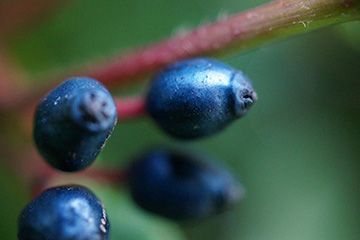
Fruits of the decorative shrub Viburnum tinus have a metallic-blue color that’s rare in the plant world. Recent work by an international team has established that the color stems from scattering of light on nanostructures made of fat molecules. [Image: R. Middleton]
Structural color is a familiar feature of the animal kingdom, visible in such archetypal examples as the peacock’s feather and the shells of iridescent beetles. In these examples and many others, the brilliant colors arise from nanostructured materials that scatter light and cause it to interfere. While rarer, structural color in plants has also been found, often tied to cellulose helicoidal architectures in the plants’ cell walls.
A multinational research team has now discovered a previously unknown way for plants to create eye-catching structural color: from layers of fat, or, more precisely, disordered multilayer lipid inclusions in the cell wall (Curr. Biol., doi: 10.1016/j.cub.2020.07.005). The lipids create a physical mulitlayer nanostructure that allows the fruits of a common landscaping shrub, Viburnum tinus, to sport a striking, metallic-blue color—signaling a nutritious treat for the birds that spread the plant’s seeds.
The team believes that the lipid mechanism could be at work in some other plants as well. And the plants’ approach might even have something to teach researchers looking at ways to create healthier, environmentally sustainable alternatives to chemical pigments.
Tantalizing fruit
The leader of the research team, University of Cambridge, U.K., physical chemist Silvia Vignolini, first became interested in V. tinus nearly a decade ago, while on a visit with the research group in Florence where she had done her Ph.D. a few years earlier. “It was kind of a random thing,” says Vignolini, who noticed the metallic-blue color of the plants’ fruit while getting a cup of coffee in an outdoor garden area. Initial measurements of the fruit, however, proved not conclusive, and she moved on to other problems.
Years later, after Vignolini had started her lab at Cambridge, one of her Ph.D. students, Rox Middleton, also grew interested in the V. tinus fruits. And they learned of other researchers that were zeroing in on the plants as well, including Michael Donaghue of Yale University, USA, and his own grad student Miranda Sinnott-Armstrong. “We decided to join forces,” says Vignolini.
Electron microscopy illuminates structure
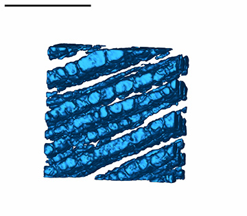
Electron and light microscopy, coupled with modeling of the structure’s optical response, established that the plant’s blue color came from a lipid-based nanostructure (shown in this schematic), rather than from blue pigments. (Scale bar: ~500 nm) [Image: M. Sinnott-Armstrong]
A key first step in sussing out V. tinus’ mechanism for structural color lay in getting solid electron microscopy (EM) to define the structure of the fruit’s cell walls. Vignolini says that her lab’s initial EM on the plant had not been very revealing. But Sinnott-Armstrong—with additional contributions from one of Vignolini’s former postdocs, Yu Ogawa of the Université Grenoble Alpes, France, and other team members—was able to use scanning and transmission EM to limn out the fruit nanostructure.
The EM study revealed—in the zone between the waxy cuticle on the fruits’ external surface that holds in moisture, and the primary cellulose cell wall—a 10-to-30-µm thick region consisting of multiple layers, each 30 to 100 nm thick, made of tiny vesicles or “blobs.” Additional EM and light-microscopy work on stained and chemically altered samples indicated the blobs were composed of lipids, specifically free fatty acids with potential nutritional value.
Modeling the optical response
The detailed work to nail down the micro-architecture and composition of the fruits’ outer layer gave the team the information it needed—such as relevant refractive-index contrasts and length scales, and the composition and characteristics of the surrounding matrix—to model the optical response, Vignolini says. In that part of the work, Middleton and her colleagues used an inverse design algorithm to establish that the detailed, disordered globular lipid structure could produce a reflectance spectrum in good agreement with the blue spectral peak measured from the fruit.
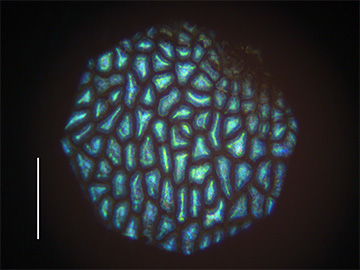
Light-microscope image of the fruit’s surface. (Scale bar: ~400 µm) [Image: R. Middleton]
The modeling thus established that the fruit’s brilliant blue is born out of light’s interaction with the lipid structure itself, with no actual blue pigment involved. Such a mechanism was a first in the study of plant structural color. “It was a surprise,” says Vignolini. “Obviously, every system does structural color with what it has available … but we had never seen [this with] lipids before.”
Message: Eat me
The strong blue spectral peak of the fruit’s measured reflectance, and in the team’s modeling of the lipid nanostructure’s optical response, lines up well with the visual systems of some birds that feed on the berries, which also appear to have a peak sensitivity in the same area. That—coupled with the fact that the blue structural color comes from lipids that themselves have nutritional value—suggests that the color could serve as an “honest signal” to birds that the fruit is well worth eating. The plant, of course, benefits by having the birds disperse the seeds embedded in the fruit.
Vignolini says that lends the story considerable biological interest. Other mechanisms that fruits use to create structural color, such as cellulose in the cell wall, “don’t have any nutritional content,” she points out. “Here, the fruit actually has nutritional content; it’s rich in lipids, which also make the optical structures. It’s saying ‘Eat me, because I have a lot of lipids and am a good food source.’”
Biomimetic-colorant potential?
Beyond illuminating biology, much of Vignolini’s work on structural color is motivated by a desire to find environmentally sustainable alternatives to hazardous chemical dyes and pigments. On that head, she suggests that something like the lipid-based approach used by V. tinus might eventually be used to healthier and safer colorants for food.
Vignolini stresses, however, that such applications are still a long way off. First, researchers will need to get a much better grasp on how the lipid nanodroplets work—and how they’re controlled and kept stable by the plant. “Before we go into the next step of a biomimetic replica of the system, which I think has potential,” she says, “the challenge is to understand how the plants do it.”
It’s a challenge to which her team is rising. The researchers believe that they have identified the same mechanism possibly at work in a number of other fruits. And a few of its members are already at work attempting to flesh out how widespread this approach to structural color might be in the plant kingdom.
In addition to researchers from Cambridge, Yale and Université Grenoble Alpes, the research team included Beverley Glover at the Cambridge Botanic Gardens and Paula Rudall of the Kew Royal Botanic Gardens, U.K.