
A new wearable probe enhances the sense of touch by imaging and quantifying the stiffness and elasticity of biological tissue. [Image: Rowan W. Sanderson, University of Western Australia]
As physicians and nurses know, there's no medical substitute for the human touch. Surgeons, for example, can tell diseased from healthy tissue by feeling for the distinguishing firmness of a small cancerous lump.
Such qualitative and subjective tactile information is hard to record or pass along to other professionals, though, so biomedical scientists have been working to measure and map such data—an imaging method known as elastography. A research team in Australia has devised the first wearable elastographic probe, incorporating an optical fiber sensor connected to a system that images the tissue deformation resulting from the surgeon's touch (Biomedical Opt. Express, doi: 10.1364/BOE.10.001760).
Beefing up elastography with OCT
In recent years, scientists have been incorporating optical coherence tomography (OCT) into their elastographic systems, creating optical coherence elastography. According to the research team from the University of Western Australia in Perth, led by first author Rowan Sanderson, OCT provides better spatial resolution and tissue-deformation sensitivity than the earlier imaging methods of elastography, ultrasound and magnetic resonance imaging.
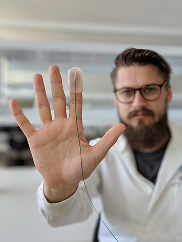
The Western Australia elastographic probe fits right onto a surgeon's gloved finger, the appendage most likely to fit into a small surgical opening such as the incision made during breast cancer removal. Specifically, the team 3-D printed a cylindrical fingertip thimble with a small channel-like extension for attaching an optical probe—a single-mode fiber spliced to a 270-μm-long graded-index fiber. Together, the fibers act as a tiny interferometer. The probe is designed for surgeons to hold and wear perpendicular to the surface of the tissue they are studying, so the light beam from the fiber probe is orthogonal to the mechanical stresses and strains on the tissue.
Testing on kangaroo tissue
The team tested the probe first on silicone “phantom” tissue that was designed with the elasticity of different types of tissue, and later on ex vivo kangaroo muscle tissue. In addition to simple 1-D touch imaging, the testers achieved 2-D scans by moving the finger-mounted probe sideways along the tissue.
The authors suggest several ways to improve their proof-of-concept device before testing on human patients. For example, the optical probe could be attached to a surgical glove for better tactile sensation and for use in a sterile operating room.