
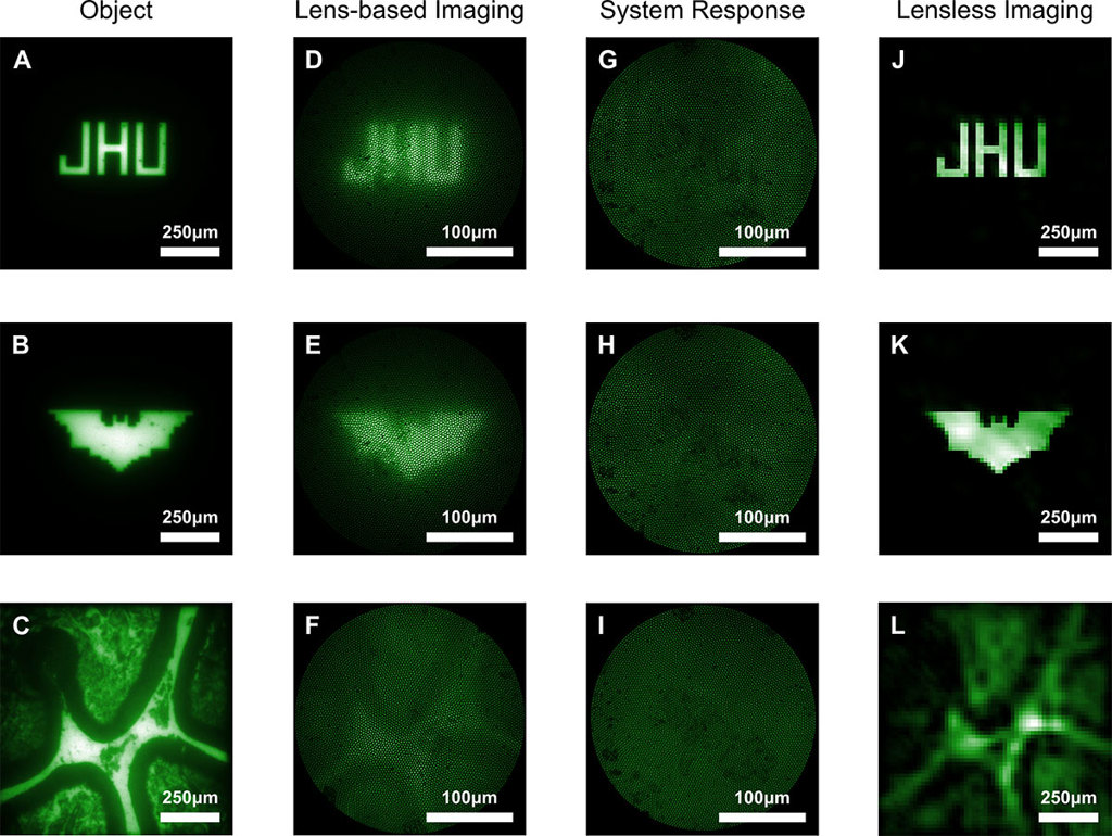
Researchers at several U.S. universities developed a lens-free microendoscope, using multicore fiber and a coded aperture, that creates images (fourth column) through computational reconstruction of the apparently random system response (third column). In proof-of-concept experiments, compared with images from a conventional lens-based microendoscope (second column), the lens-free, computational microendoscope provided a clearer picture of the original objects (shown, as imaged by a bulk microscope, in the first column). [Image: Mark Foster] [Enlarge image]
To investigate the detailed inner workings of the brain, neuroscientists insert optical endoscopes in living animals to observe neural activity with single-cell resolution—but such devices typically sacrifice image quality for a small size. Now, a team of U.S. researchers has created a lens-free, ultrathin microendoscope the width of only a few human hairs, with a broad field of view (Sci. Adv., doi: 10.1126/sciadv.aaw5595).
Measuring neural activity
Mammalian brain tissue strongly scatters light, which proves challenging for non-invasive optical imaging techniques. Methods like two-photon microscopy can easily investigate a superficial layer of the brain, up to about one millimeter in depth. But going any further requires physically inserting a probe. Endoscopes should be miniaturized as much as possible to prevent tissue damage that could adversely affect the animal—and, consequently, any experimental results.
“We’re interested in understanding how neural circuitry works, which is the reason why we were looking at making these really small microendoscopes,” said Mark Foster of John Hopkins University, the new study’s corresponding author. “When you do so, you encounter a problem where the smaller you make it, the less image information you can collect.”
A different approach
Traditional microendoscopes mechanically scan an optical fiber with a microlens to acquire each image pixel of a scene. These devices have a reduced field of view, limited by the deflection angle of the scanner, and the lenses add bulkiness to their overall size. Microendoscopes that use a multicore fiber or fiber bundles have poor image quality due to crosstalk between fiber cores and are difficult to miniaturize.
Foster and his colleagues took the novel approach, using multicore fiber to create a lens-free microendoscope. Instead of the usual distal lens, the researchers inserted a pseudorandom patterned mask, known as a coded aperture, at the end of the optical fiber. A CCD camera at the other end of the fiber records a linear combination of light emitted from various points within the scene. A computational algorithm then takes these system responses, which look like a random intensity fluctuation to the naked eye, and reconstructs an image of the object.
Superior image quality
The team thoroughly calibrated and tested the system, starting with projections and then moving onto objects. Compared with a lens-based system, the lens-free microendoscope produced images with superior image quality and was capable of resolving features only 14 μm in size. The device also performed computational refocusing of objects located 1.5 mm apart in depth with a single snapshot and achieved color imaging without any additional components.
“The system does everything we wanted in a simple, compact package,” said Foster. “The next step is to transition the system from widefield imaging to fluorescence imaging and eventually move onto in vivo imaging.”
In addition to researchers at Johns Hopkins University, the team also included scientists from Boston University, Massachusetts Institute of Technology, and Harvard University.