
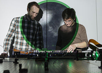
The projection behind lead authors Sergey Turtaev (left) and Ivo T. Leite (right) shows an image of neurons obtained deep inside the brain via a single multimode fiber. [Image: Sven Doering / Liebniz-Institute of Photonic Technology, Germany]
Researchers from Germany and the United Kingdom have developed an endoscopy system for deep-brain in vivo high-resolution fluorescence imaging at the tip of a multimode optical fiber. The imaging system uses new methods for holographic control of light propagation in complex media, such as the brain, to increase imaging speed and improve image quality (Light Sci. App., doi: 10.1038/s41377-018-0094-x). The researchers successfully demonstrated their minimally invasive imaging system on deep-brain regions in live mouse models.
The team, led by Tomas Cizmar of the Liebniz-Institute of Photonic Technology, Germany, and Nathalie Rochefort from the University of Edinburgh, U.K., believes that the imaging system could enable scientists to learn more about how neurons function in the brains of awake and behaving animals without harming the surrounding tissue. According to the researchers, the technology could someday prove useful for research into sensory perception, memory formation and brain diseases like Alzheimer’s.
Holographic endoscopy
Current endoscopy methods, which are based on either bundled optical fibers or graded-index lenses, are capable of subcellular imaging but are not ideal for use in the brain because their relatively bulky size can cause damage to the surrounding tissues. External imaging methods, like magnetic resonance imaging (MRI), are non-invasive but not powerful enough to capture the small neuronal structures in the brain.
Advances in computer-controlled holographic modulators, says the German–U.K. team, may eliminate this trade-off between system size and imaging resolution by making high-resolution fluorescence imaging possible through a very thin multimode optical fiber (MMF). The new holographic endoscope system contains a digital micromirror device (DMD), with binary-amplitude gratings based on the Lee hologram approach, that behaves as a spatial light modulator. The DMD is in the system’s beam-shaping module and holographically controls the phase and amplitude of light propagating through the MMF and brain tissue, allowing for higher quality images and faster acquisition speeds.
Putting it all together
The MMF-based endoscopy system consists of an endoscopic probe, a laser, a beam-shaping module, a calibration module and a sample module. The probe is a standard 50-µm-diameter MMF. The light source is a single-frequency, diode-pumped solid-state laser emitting at 532 nm that can provide a continuous-wave or linearly polarized beam. The beam-shaping module contains the DMD to holographically control the phase of the optical fields coupled into the fiber.
With the calibration module in place, the DMD in the beam-shaping module helps the system characterize the laser light that propagates through the system’s optical path at various focal planes behind the end of the MMF. This characterization is referred to as the system’s transmission matrix (TM). It takes about two minutes for the system to generate a TM and calculate about 7,000 phase-modulation patterns for all foci across the MMF’s 50-µm field of view (FOV). Once the calibration is complete and the data are saved to the DMD’s memory chip, the calibration module can be replaced with the sample module.
The researcher say that with the sample module in place, the system can achieve 1-µm resolution across a 50-µm FOV, creating 7-kilopixel images at a speed of 3.5 frames per second.
Deep-brain in vivo imaging
The researchers demonstrated the MMF-based endoscopy system on live, anesthetized mouse models with inhibitory neurons in the brain labeled with a red fluorescent marker. They reportedly captured images of individual nerve cells and neuronal processes—objects with diameters ranging from 10 µm to 2 µm—in the brain’s visual cortex and the hippocampus, with a minimal impact on the structure and function of the surrounding brain areas. The system was also able to record, over time, individual red blood cells from a burst blood vessel in the visual cortex of the mouse brain.
The team says that its research could “pave the way” for new approaches to the use of multiphoton, super-resolution and light-sheet microscopy for many biomedical applications.