
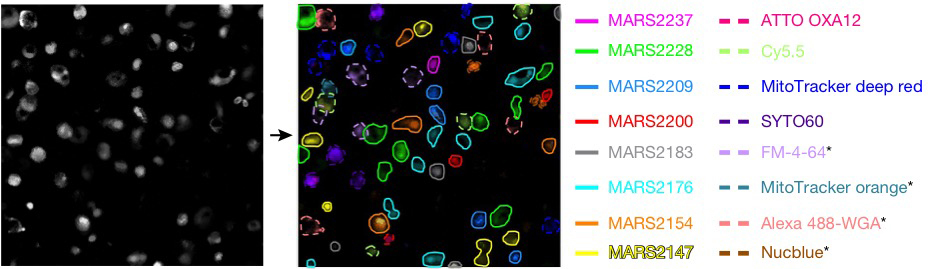
Demonstration of super-multiplex optical microscopy. Left: Pre-stained cells. Right: Live-cell imaging with eight MARS dyes (solid), four vibrational dyes in the Raman fingerprint region (dashed), and four fluorescent dyes (asterisk). [Image: L. Wei et al. Nature, doi: 10.1038/nature22051] [Enlarge image]
Researchers from Columbia University, USA, say proof-of-principle demonstrations of their new super-multiplex vibrational imaging platform could eventually expand the current, limited five-color palette available with fluorescence microscopy by nearly five times, to 24 colors (Nature, doi:10.1038/nature22051).With this new platform, the team says researchers could eventually view 24 different sub-cellular structures simultaneously in a single sample, with higher specificity and sensitivity than traditional fluorescence and Raman microscopy techniques.
The Columbia team’s platform combines two synergistic pieces—a more sensitive form of Raman scattering microscopy instrumentation and several new near-infrared dye molecules. The researchers believe this new platform, called electronic pre-resonance stimulated Raman scattering (epr-SRS) microscopy, will eventually contribute to finding new therapies to treat and cure diseases.
Improving sensitivity and specificity
Team leader Wei Min says the detection sensitivity of the group’s epr-SRS microscopy platform is several orders of magnitude better than conventional Raman microscopy because it “harnesses the stimulated emission process to amplify the otherwise weak vibrational transition.” The team estimates that epr-SRS can detect molecular-structure concentrations as low as 30, compared with the millions of structures needed to generate a signal from conventional Raman microscopy.
The researchers conducted a specificity proof-of-principle demonstration with human epithelial cells (HeLa cells) fluorescently tagged with ATTO740 dye for a specific metabolic target. They found that the ATTO740 signal disappeared if the pump laser’s wavelength was off by 2 nm. This, according to the researchers, indicates chemical specificity beyond that of normal fluorescence microscopy. The team also tested the platform’s versatility by confirming its ability to image genetically encoded proteins in live cells, organelle targets and chemical drugs; as well as its use in immunolabeling and small-molecule-based functional assays.
Expanding the dye palette
After demonstrating the epr-SRS platform’s specificity and sensitivity, the researchers expanded the vibrational (i.e., Raman fingerprint region) palette of dyes to 14 resolvable reporter dye molecules between 650 and 800 nm, which they named the Manhattan Raman scattering (MARS) dyes. By combining the MARS dyes with six commercially available dyes in the Raman fingerprint region and four fluorescent dyes in the visible region, they created a 24-color palette for optical super-multiplexing.
Color palette proof-of-principle demonstrations were conducted with HeLa cells and brain tissue cells under different physiological and pathological conditions. The cells were separately stained with 16 different dyes before being mixed together and placed under a microscope. The 16-color image shown above [enlarged version here] resulted from the sequential imaging of a cell sample over 15 minutes. Additional proof-of-principle demonstrations of live brain-cell super-multiplexing showed successful eight-color imaging of metabolic activity, DNA replication and protein synthesis.
Increasingly colorful
According to a press release, the researchers believe that there’s potential for even further color expansion. In the future, the researchers hope to be able to image structures in other tissue types in real time; more colors aid the study of how multiple cells interact and function simultaneously. The team would also like to be able to observe how cells interact with each other in both healthy and diseased tissues, in hopes of identifying ways to target structures for more precise therapies.