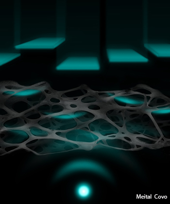
An artist’s rendering of a shaped femtosecond pulse wavefront focusing through a scattering tissue.
Obtaining clear images of objects through thick, turbid layers, like biological tissues, is crucial in medicine and biological research. However, it poses serious challenges because light scattering through the complex media distorts the images produced. Now, a team of researchers from Israel and China has reportedly demonstrated a new technique that uses optical nonlinearities to form a diffraction-limited focus through highly scattering biological samples with visually opaque layers (Optica, doi: 10.1364/optica.1.000170).
The team integrated adaptive optics—via a high-resolution wavefront shaping device—into a standard laser scanning two-photon microscope, to help focus an image of a single point behind a scattering layer. Traditionally, adaptive optics requires a stable reference point, or “guide star,” in the same area as the object of focus. The guide star provides direct feedback on the distortion of the signal, which allows for adjustment and correction of the wavefront to compensate for scattering. But this is invasive and impractical for many biological applications, as the guide star has to be physically inserted into the sample at the target position to be imaged.
As an alternative method, the researchers exploited the nonlinear nature of two-photon fluorescence to “pre-correct” the laser beam, according to co-author Ori Katz of the Weizmann Institute of Science, Rehovot, Israel. In their setup, 100 fs laser light pulses pass through a phase-only spatial light modulator (SLM) before entering the sample, and the total backward-scattered nonlinear fluorescence signal is epi-detected using an integrating detector. Then, that total nonlinear signal—rather than the image actually formed by the distribution of the fluorescence—is optimized using a genetic algorithm that iteratively tweaks the SLM phase pattern.
The probe light is thus adaptively shaped into a beam that penetrates the obscuring layer and can tightly focus on target behind the layer that’s less than one micron in diameter—without previous knowledge of the target’s precise location. The authors believe that this technique should be adaptable to most nonlinear microscopy techniques, including two- and three-photon fluorescence, second- and third-harmonic generation, four-wave-mixing, and coherent anti-Stokes Raman scattering. Further research will include shortening the time required to achieve the correct focus.
{^media|(width)700|(height)450|(ext).mov|(url)https://opnmedia.blob.core.windows.net/media/opn/media/images/homepage/multimedia/170.avi^}
