
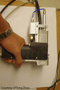
A photo of the handheld probe.
Melanoma is the deadliest of all skin cancers, and its successful treatment depends on early detection and characterization of tumors—including their thickness. But current approaches to measuring tumors in the clinic all have shortcomings in depth penetration or 3-D resolution. Now, researchers from Washington University in St. Louis, U.S.A., have introduced a handheld probe that uses lasers and sound waves to delineate the boundaries and measure the thickness and volume of melanoma tumors (Opt. Lett., doi: 10.1364/OL.39.004731). According to the team, the instrument is the first that can be used directly on a patient to accurately measure the depth of a melanoma tumor in the skin.
The probe contains a motor, translation stage, laser, ultrasonic transducer and optical fibers and can be used directly on the patient’s skin. First, the probe lights up an area of the skin with short laser pulses. After the skin absorbs the light, ultrasonic waves are induced thermoelastically through the photoacoustic effect. Since most melanomas are heavily pigmented compared to the surrounding skin, they can be imaged with photoacoustic microscopy with high contrast. And since acoustic scattering through skin is low compared to light scattering, users can obtain high-resolution images much deeper than with optics-based instruments.
The team tested the handheld probe on artificial black gelatin tumors and melanoma tumors in mice. With their device, they were able to measure tumor depths up to 4.1 mm in gelatin tumors and 3.7 mm in mouse tumors, outperforming optical methods that often miss the 2 mm to 4 mm range needed to stage melanoma tumors. Thickness and staging are important values because they directly relate to the chances of a patient developing metastatic disease, which increases disease mortality.
The researchers also found that their device could deliver light above and around the tumor, which provides data to create a 3-D image of the tumor and to calculate its volume. A 3-D image could improve the accuracy of surgical tumor removal; volumetric measurements could give physicians insight into how tumor volume relates to treatment outcomes.
Imaging speed is currently a limiting factor for the device—the probe’s 10 Hz laser takes ten seconds to scan a 10-mm length with a step size of 100 µm. However, a pump laser system enhancement may increase speed for clinical use. The researchers are conducting tests with human patients and are looking ahead to clinical trials of the device.