
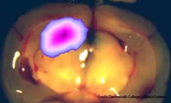
Fluorescent in vivo image of exposed mouse brain following resection and exposure of the brain tumor (false colors).
Surgeons want to use emerging fluorescence-based techniques to guide their hand when resecting diffuse tumors, but current systems work only when all the overhead lights in the operating room are shut off—a practice surgical teams dislike. Researchers at Dartmouth College (U.S.A.) have devised a pulsed-light imaging technique that delineates tumor cells while the room lights remain on (Opt. Lett. 38, 3249).
By exciting fluorophores in mouse tumors with pulsed lighting instead of a continuous source, and using a synchronized time-gated detector for imaging, the proof-of-concept system reduces the contribution of background lighting to the desired signal. OSA Fellow Brian W. Pogue says that one of his co-authors, neurosurgeon David W. Roberts, likens existing fluorescence-guided surgical setups to “trying to operate under the moonlight.”
In some cases, cancer cells preferentially absorb the precursors to fluorescent porphyrins and generate more of the porphyrins than surrounding tissue, says Kristian Sexton, a graduate student at Dartmouth's Thayer School of Engineering. Physicians want to exploit this tendency to aid in removing tumors that are not well-circumscribed.
Pogue, Sexton and their colleagues in the Dartmouth engineering and medical schools illuminated the surgical field with four 630-nm high-instantaneous-brightness LEDs. The diodes emit 500-μs pulses at a 50-Hz rate, so the illumination is off about 1,000 X more than it is on, and each pulse delivers about 8,000 mW per cm2—about 40 X the power that could be safely delivered by continuous lighting. For lighting a broad area, LEDs are less costly per photon than lasers, according to Pogue.
During a procedure, the surgeon looks at a monitor and sees the false-color fluorescence signature from the microscopic imaging system superimposed over full-color video of the surgical field. Tests with brain surgery on mice showed that the experimental system's sensitivity under regular room lighting is about an order of magnitude better than that of a commercially available system used in the dark.
The team will use a camera with higher resolution and a better lens in future iterations of the experiment. The team is working on a dual-bandwidth system to pick up fluorescent signals from multiple tracers.