
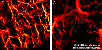
Two 2x2 mm areas showing (a) a healthy network of blood, and (b) blood vessels supplying a basal cell carcinoma. Note the branching pattern and abnormally large vessels.
A non-invasive optical technique that maps tiny blood vessels under the skin successfully detected skin cancer in a small study. Rainer Leitgeb and others from Medical University Vienna (MUW, Austria) and the Ludwig-Maximilians University (Munich, Germany) used optical coherence tomography (OCT) to image microvasculature, and showed that basal cell carcinoma, inflammation and healthy skin show distinct characteristics (Biomed. Opt. Express, doi:10.1364/BOE.3.002636 ).
Many cancers alter the growth of blood vessels, but these changes are not always obvious to human sight. OCT, which is analogous to ultrasound (but at a much finer scale), images into tissue, and has been used in the past to image cells. Instead, the researchers developed a high-speed OCT to image microscopic blood vessels near the surface of skin. They tested subjects with a healthy human palm, allergy-induced eczema on a forearm, dermatitis on a forehead and two cases of basal cell carcinoma (the most common type of skin cancer) on faces. They found that the network of vessels supplying blood to the tested lesions show patterns that are significantly different from those of healthy skin.
The OCT images had to be acquired quickly to avoid artifacts of patients moving. The researchers developed a high-speed Fourier domain mode-locked laser emitting at 1310 nm as a fiber-coupled source. The near-IR wavelength is scattered less than visible wavelengths, improving the signal-to-noise ratio and allowing volumetric imaging over a depth range of about 1 mm. The system also allows researchers to choose either a comparatively large lateral resolution or a larger depth range. The output was a Bessel beam, shaped by a lens and mask into a ring, which (like other Bessel beams) can reconstruct its shape, thus reducing the effect of hair or other occlusions.
Compared to healthy skin, they found that inflamed skin had dilated and leaky vessels, while cancerous areas showed a denser, disorganized network with large vessels close to the surface. “The condition of the vascular network carries important information on tissue health and its nutrition,” says Leitgeb. “Currently, the value of this information is not utilized to its full extent.”
A more-developed non-invasive diagnostic method based on this technology could guide dermatologists to the most likely spots for cutting out tissue for biopsies—and might eventually reduce the number of biopsies needed.