
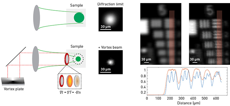
 Left: Schematic of vortex-assisted focusing. Center: Experimental comparison between a diffraction-limited spot and one obtained via vortex assisted focusing. Right: Scanning microscopy image of USAF resolution chart without (left) and with (right) the vortex plate. Image resolution is enhanced, as can be quantitatively seen from the line plot of the marked region. [Figure is reproduced with permission from ref. 4]
Focusing light beyond the diffraction limit is among the greatest challenges in optical sciences, and would revolutionize many optical technologies from imaging and optical trapping to light delivery and photomedicine. To achieve sub-diffraction focusing, metamaterials,1 materials with engineered scattering properties2 or attached microspheres3 have been proposed. Recently, we introduced a way to achieve a physical focus beyond the diffraction limit without additional optical elements or exogenous labels, using only light–matter interactions.4
To achieve tight focusing of a probe beam (green beam in accompanying diagram), negligibly absorbed by the sample, our technique uses an auxiliary beam (red) of different wavelength, which is strongly absorbed by the sample. In water-based solutions, the temperature dependence of the refractive index is negative (dn/dt < 0). Therefore, the auxiliary beam is shaped by a vortex phase plate to generate transient absorption in the shape of a donut—that is, a ring with a diffraction-limited void at the center.
This heat profile induces a refractive-index gradient at the waist of the focusing lens, with highest index at the center of the ring and with the index monotonically decreasing toward the periphery. This process effectively sculpts a transient converging microlens with diameter equal to the optical diffraction limit. Thus, when the probe beam passes through the sculpted microlens (either collimated or focused), it will converge to a tighter focal point than what is allowed by the optical system, as we confirmed experimentally.
Because of the short pulse duration needed to sculpt the heat profile, the exposure flux of our imaging modality is below the damage threshold of tissues, making it suitable for biological applications. The smaller focal point can be also used as a scanning probe for imaging at superior resolution without any labeling process or material insertion, providing high flexibility in the optical configuration.
Researchers
Eitan Edrei and Giuliano Scarcelli, University of Maryland, College Park, MD, USA
References
1. N. Fang. Science 308, 534-537 (2005).
2. M. Jang et al. Nat. Photon. 12, 84 (2018).
3. Z.B. Wang et al. Nat. Commun. 2, 218 (2011).
4. E. Edrei and G. Scarcelli. ACS Photon. 7, 914 (2020).