
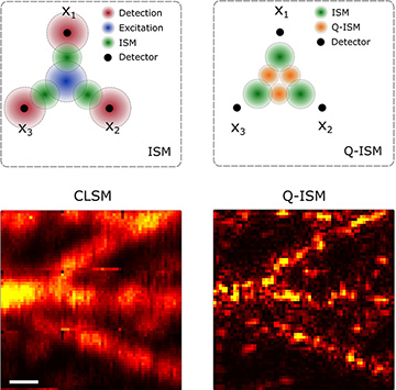
 Top: In conventional ISM (left), the PSF for each detector (green circles) is a product of the excitation laser beam profile (blue circle) and the detection probability distribution (red circles). In Q–ISM (right), the effective PSF for a detector pair (orange circles) is a product of the two ISM PSFs (green circles). Bottom: Confocal laser scanning microscopy (left) and Fourier-reweighted Q–ISM (right) images showing microtubules in a fixed 3T3 cell labeled with fluorescent quantum dots; scale bar = 0.5 μm. [Adapted with permission from reference 3.] [Enlarge figure]
Light microscopy, one of optical science’s oldest applications, has always been strongly driven by technological advances. The recent evolution of quantum technologies, relying on advanced generation and detection methods for quantum states of light, has stimulated new approaches to overcome microscopy’s classical limitations. This year, we reported one such approach, which uses quantum correlations to improve image-scanning microscopy (ISM).
Generating entangled photon pairs and measuring their correlation after interacting with a sample have previously enabled both super-resolution and sub-shot-noise phase imaging. However, since quantum states of light are sensitive to loss through scattering and absorption in biological samples, their application in life science imaging has proved very challenging.
An alternative approach is to label the sample with markers that generate a quantum state of light. A dye molecule or a semiconductor nanocrystal emits a single photon at a time, giving rise to the quantum-optical phenomenon of photon antibunching. While measuring the resulting anti-correlation in the detection of photons can yield a super-resolved image on its own, it also has further potential in enhancing already established super-resolution methods.
Our work combines the measurement of quantum correlations with ISM. In standard ISM, a single count requires both an excitation by a focused laser beam and a detection of an emitted photon by one of the detectors in a small array. Therefore, a scanned image’s effective point spread function (PSF) from each detector is a product of the laser excitation profile and the detection PSF. The sum of (properly) shifted images from the multiple pixels produces a super-resolved image, without sacrificing the signal levels.1,2
In our quantum version, Q–ISM,3 a signal count is interpreted as the lack of a simultaneous photon-pair detection. Such a “missing” event corresponds to the absorption and detection of two photons. Therefore, the resulting PSF is a fourth power of the original PSF, with up to a fourfold improvement beyond the diffraction limit. Applying Q–ISM, we obtained super-resolved images of fixed cells whose contrast solely relies on a quantum-optical effect.
Noting that the quantum correlation image is acquired in parallel with the classical ISM image, we believe that there is plenty of room to improve classical imaging modalities through measurement of a complementary quantum contrast.
Researchers
Ron Tenne, Uri Rossman, Batel Rephael, Yaron Silberberg and Dan Oron, Weizmann Institute of Science, Rehovot, Israel
Yonatan Israel, Weizmann Institute of Science, Rehovot, Israel, and Stanford University, Stanford, Calif., USA
Alexander Krupinski-Ptaszek and Radek Lapkiewicz, University of Warsaw, Warsaw, Poland
References
1. C.J.R. Sheppard. Optik (Stuttgart) 80, 53 (1988).
2. C.B. Müller and J. Enderlein. Phys. Rev. Lett. 104, 1 (2010).
3. R. Tenne et al. Nat. Photon. 13, 116 (2019).