
Feature
Polarization-Resolved Microscopy in the Life Sciences
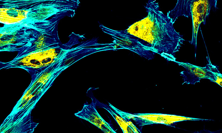
Using light polarization as an additional degree of freedom in microscopy can provide new insights into molecular organization.
 [Getty Images]
Optical microscopy offers invaluable access to biomolecular processes at sub-micron and even single-molecule scales. It occupies a unique position between techniques such as magnetic resonance imaging and ultrasound, which provide deep penetration but low spatial resolution, and others such as electron microscopy and X-ray diffraction, which yield sub-nanometer resolution but cannot image live organisms. Optical microscopes can image live organisms and tissues at relatively high spatial resolution (around 200 nm), large fields of view (up to millimeters) and reasonable penetration depths (a few hundreds of microns). And optics can specifically address biomolecules such as proteins, DNA and lipids in real time with single-molecule sensitivity using fluorescence labelling, or even via label-free imaging by taking advantage of nonlinear optical effects in tissues.
…Log in or become a member to view the full text of this article.
This article may be available for purchase via the search at Optica Publishing Group.
Optica Members get the full text of Optics & Photonics News, plus a variety of other member benefits.