
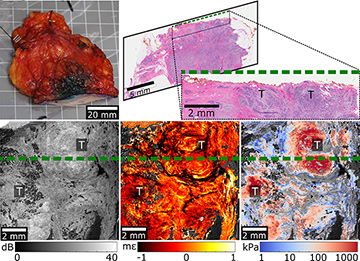
 QME of a BCS specimen with an involved margin (also see accompanying video.) Top: Specimen photograph (left) and histology (right). Bottom: En face OCT (left), strain (center) and elasticity (right) images. The green line indicates the location of the histology slide, which is orthogonal to en face images. T, invasive tumor. [Enlarge figure]
Many breast cancer surgeons still rely on their sense of touch—manual palpation—for guidance during breast-conserving surgery (BCS), by feeling for changes in mechanical properties associated with tumor. However, manual palpation is subjective and offers an inherently low resolution.1 This is a main contributing factor to high re-excision rates, currently one-in-four in BCS.2 Reducing those rates, and thus improving patient outcomes, will require more accurate intraoperative tools for assessing tumor margins.
Quantitative micro-elastography (QME) is an emerging imaging technique that aims to transform the resolution of manual palpation to the micro-scale. Based on optical coherence tomography (OCT), QME measures depth-resolved mechanical parameters—that is, strain and elasticity—of soft tissues, thereby providing mechanical contrast which complements the optical contrast from OCT.3
This year, we presented a feasibility study in which we incorporated QME into a wide-field imaging system to increase the contrast between tumor and normal dense tissue.4 In the study, we imaged 13 specimens from patients undergoing mastectomy at Fiona Stanley Hospital, Western Australia. We presented wide-field (46×46-mm) strain and elasticity images, alongside OCT images, all generated from a single QME dataset acquired in 10 minutes.
The study demonstrated that the triple contrast provided by elasticity, strain and OCT improves visualization of tumor, in particular, high-grade ductal carcinoma in situ and highly cellular invasive ductal carcinoma. The study also demonstrated a reduction in imaging artifacts and an increase of overall tissue area assessed compared to a related micro-elastography technique5—both factors that can potentially increase diagnostic accuracy.
QME images of an intact BCS specimen (see figure), mimicking the clinical scenario for tumor margin assessment, show invasive ductal carcinoma within 300 µm of the margin. The tumor can be delineated by the high elasticity as well as by the heterogeneous OCT and strain signal in en face images.
The combined evaluation of mechanical and optical contrast provided by QME has enabled us to visualize a broader range of tumors than is possible with OCT alone. The methods developed in the study will enable us to move the clinical development of QME forward. We believe that this technique, if deployed intraoperatively, has the potential to reduce re-excision rates by enabling surgeons to detect tumors that may otherwise be missed using manual palpation alone.
Researchers
Wes M. Allen, Kelsey M. Kennedy, Qi Fang, Lixin Chin, Andrea Curatolo, Renate Zilkens and Brendan
F. Kennedy, Harry Perkins Institute of Medical Research and The University of Western Australia, Perth, Australia
Synn Lynn Chin and Bruce Latham, Fiona Stanley Hospital, Murdoch, Australia
Benjamin F. Dessauvagie, The University of Western Australia and Fiona Stanley Hospital
Christobel M. Saunders, The University of Western Australia, Fiona Stanley Hospital and Royal Perth Hospital
References
1. R.G. Pleijhuis et al. Ann. Surg. Oncol. 16, 2717 (2009).
2. H. Ballal et al. ANZ J. Surg. 85, 540 (2015).
3. K.M. Kennedy et al. Sci. Rep. 5, 15538 (2015).
4. W.M. Allen et al. Biomed. Opt. Express 9, 1082 (2018).
5. B.F. Kennedy et al. Cancer Res. 75, 3236 (2015).