
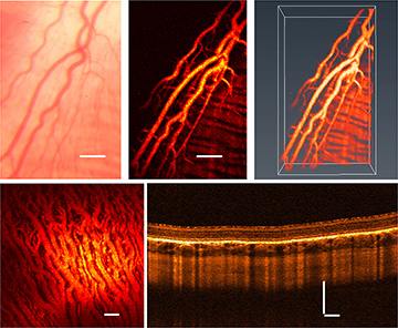
 Top left: Fundus photo of retinal vessels of a living rabbit. Top center and right: PAM image of the same area and its 3-D rendering. Bottom: PAM image of choroidal vessels (left) and an OCT B-scan (right). Scale bar: 200 µm. [Enlarge figure]
Vision impairment is a major public disability, affecting more than 285 million people worldwide. Evidence shows that it is preventable or curable in 80 percent of cases if properly diagnosed and managed. The eye is optically transparent in health, and thus optical imaging plays a pivotal role in this important health issue. This year, we developed a high-resolution photoacoustic microscopy technique for imaging of the eye.
Currently available techniques for optically imaging the eye mainly include fluorescein angiography (FA), indocyanine green angiography (ICGA), optical coherence tomography (OCT) and OCT angiography (OCTA). FA and ICGA require intravenous injection of exogenous fluorescence dyes to visualize retinal and choroidal vasculature, but that invasive procedure can cause adverse reactions in up to 10 percent of patients. OCT and OCTA can visualize anatomical structure and vascular networks of the retina, but give limited contrast for evaluating molecular changes. In addition, the blood supply for the visual photoreceptors comes from a deeper vascular supply called the choroid, at a depth to which OCT has difficulty penetrating. It’s thus essential to develop high-resolution advanced retinal and choroidal imaging technology with a greater penetration depth.
Photoacoustic imaging, which converts light energy into sound waves, is an emerging technique that has found application in numerous biomedical fields. Eyes are an ideal system, and photoacoustic imaging of the eye has attracted substantial interest.1
In contrast to conventional imaging technologies, photoacoustic imaging is based on the optical-absorption contrast, and can provide unique pathophysiological information for the eye as well as a deeper penetration depth. Most work to date has focused on imaging small animals, such as mice, the eyes of which are only 12 percent of the length of the human eye. Since the photoacoustic signal attenuates quickly with propagation distance, photoacoustic imaging of larger eyes is particularly challenging.
This year, our group developed high-resolution photoacoustic microscopy (PAM) imaging (4.1 µm lateral resolution) of the retina and choroid for the larger eyes of organisms like rabbits, which have an eye length 80 percent of that of humans. By exploiting optical-absorption and optical-scattering contrasts of ocular tissue at the same time, we successfully achieved label-free choroidal and retinal imaging in living rabbits using novel integrated PAM and OCT.2,3 Our results showed that PAM could noninvasively resolve retinal and choroidal blood vessels at single-vessel resolution using a safe laser exposure in living rabbits, and while OCT visualized the anatomic layers.
We believe that the work holds great promise in basic research and clinical practice in ophthalmology, by providing optical-absorption-based, high-resolution images of deeper eye structures.
Researchers
Chao Tian and Yannis M. Paulus, University of Michigan, Ann Arbor, Mich., USA
References
1. W. Liu and H.F. Zhang. Photoacoustics 4, 112 (2016).
2. C. Tian et al. Opt. Express 25, 15947 (2017).
3. C. Tian et al. J. Vis. Exp., e57135 (2018).