
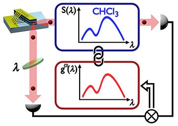
 Ghost spectroscopy demonstration experiment exploiting spectrally correlated photons from a superluminescent diode, within a real-world absorption spectroscopy experiment on liquid chloroform (CHCl3). The ghost spectrum, measured in terms of the spectral second-order correlation coefficient g(2)(λ) (red data), reproduces the standard absorption spectrum S(λ) of chloroform (blue data). [Enlarge figure]
The quantum imaging technique known as ghost imaging (GI) exploits photon correlations for image construction. One photon of an (entangled) pair interacts with the object, and the experimentally determined correlation with the second photon yields the image. The intensity autocorrelation or second-order correlation is thus transferred into a spatial image of the object—the “ghost” image. GI was first achieved in 1995 with entangled “twin” photons generated by spontaneous parametric down-conversion.1 Classical GI, first experimentally demonstrated in 2004 with pseudo-thermal light,2 was later achieved even with a neon discharge lamp and, particularly interesting, also with semiconductor-based emitters.3
This year, we succeeded in transferring the concept of classical spatial correlations into the spectral domain, thus realizing, for the first time, a ghost spectroscopy experiment with classical thermal photons.4 We exploited wavelength–wavelength correlations of broadband amplified spontaneous emission (BB-ASE) light emitted by miniaturized, optoelectronic, semiconductor-based quantum dot superluminescent diodes (SLDs). In the spirit of the famous Hanbury Brown and Twiss experiment,5 we showed that this light exhibits a spectral second-order correlation coefficient of two and, thus, spectral photon bunching—one of the key requirements of ghost spectroscopy.
To demonstrate this light source’s applicability to ghost spectroscopy, we conceived a “real-world” absorption spectroscopy experiment.4 BB-ASE light from an SLD at a central wavelength of 1228 nm exhibiting the spectral second-order correlations was divided into a wavelength-correlated object and a reference beam, respectively. The object beam was transmitted through a sample cell filled with liquid chloroform (CHCl3). Its transmitted intensity was measured spectrally integrated—that is, not spectrally resolved—whereas the reference beam, which had never “seen” the sample, was spectrally resolved and detected by tunable interference filters.
Therefore, neither beam, alone, contained any direct spectral information on the sample. Correlating the intensities of reference and object beam by means of a two-photon-absorption photomultiplier, however, yielded the second-order correlation coefficient g(2)(λ) that carried the ghost spectrum, which clearly exhibited all the characteristic absorption fingerprints S(λ) of chloroform at 1214 nm. The ghost absorption spectrum obtained in this manner is thus the wavelength-domain analogue of a ghost image in the spatial domain.
This first demonstration of ghost spectroscopy with classical thermal light, in analogy with GI, closes a gap in experimental photon correlation modalities. We expect that the result will further fertilize and stimulate the ghost modality idea toward further applications in chemistry, physics and engineering.
Researchers
Patrick Janassek, Sébastien Blumenstein and Wolfgang Elsäßer, Technische Universität Darmstadt, Germany
References
1. T.B. Pittmann et al. Phys. Rev. A 52, R3429 (1995).
2. F. Ferri et al. Phys. Rev. Lett. 94, 183602 (2005).
3. S. Hartmann et al. Sci. Rep. 7, 41866 (2017).
4. P. Janassek et al. Phys. Rev. Appl. 9, 021001 (2018).
5. R. Hanbury Brown and R.Q. Twiss. Nature 178, 1046 (1956).