
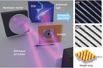
 Left: Soft-X-ray microscope design achieves a resolution better than the illuminating wavelength. Right: Comparison between a scanning electron (SEM) microscope image and the coherent lensless diffraction soft-X-ray image (CDI) (note that the contrast is reversed, as expected), as well as a height map of the sample.
Visible-light microscopes can acquire stunning images at high spatial resolution, using near-perfect lenses that focus light to spot sizes smaller than the wavelength—to the Abbe limit of roughly λ/2 NA, where NA is the numerical aperture. Soft-X-ray (SXR) light has wavelengths 10 to 100 times shorter than visible light. Thus, it should be possible to design a powerful microscope that can see structures too small or too opaque to be seen with visible light.
Despite these advantages, however, progress in extreme-UV (EUV) and SXR microscopy during its 60-year history has been limited by two major challenges: a lack of bright coherent sources in the EUV/SXR regions, and the fact that diffractive (zone-plate) X-ray optics used for image formation are lossy, imperfect and severely limited in spatial resolution, to around 10λ. As a result, until the 2010s, the best spatial resolution achieved in SXR with test samples was around 12 nm (using 2-nm synchrotron beams), while for real-world samples, the spatial resolution was typically 25 to 30 nm.
Fortunately, these challenges have been addressed in recent years. Tabletop coherent EUV/SXR beams are now possible using high-harmonic generation (HHG).1,2 In addition, a new generation of powerful coherent-diffractive-imaging (CDI) techniques is removing the resolution limits imposed by traditional X-ray microscopy, by replacing lossy and imperfect X-ray optics with powerful iterative phase retrieval algorithms.3
In particular, a CDI technique called ptychography has yielded exceptional results in recent years.4 In ptychography, the scatter patterns from neighboring and overlapping regions are collected, and the resulting redundancy in the diffraction data enables robust image reconstruction with exquisite phase contrast.
Recent collaborative work by JILA and KMLabs has extended ptychographic techniques to demonstrate the first subwavelength-resolution imaging at short wavelengths.5 We achieved high-quality imaging of an extended sample with a spatial resolution of 0.9λ, and also demonstrated high-fidelity, full-field, quantitative imaging of near-periodic objects.
Both the 13.5-nm wavelength used in this work and the ability to reliably image near-periodic objects are technologically relevant for the development of next-generation EUV lithography, nanoelectronics, data storage and self-assembled nanostructures, as well as functional imaging of nano-enhanced devices. Moreover, because HHG produces exceedingly short (roughly 10–15 sec) bursts of light, it can also be used to make stroboscopic movies to observe how the nanoworld functions.
Researchers
Michael Tanksalvala, Christina Porter, Dennis Gardner, Xiaoshi Zhang, Elisabeth Shanblatt, Benjamin Galloway, Robert Karl Jr., Charles Bevis, Giulia Mancini, Daniel Adams, Henry Kapteyn and Margaret Murnane, JILA, University of Colorado and KMLabs, Boulder, Colo., USA
References
1. A. Rundquist et al. Science 280, 1412 (1998).
2. R.A. Bartels et al. Science 297, 376 (2002).
3. J. Miao at al. Science 348, 530 (2015).
4. J.M. Rodenburg. Adv. Img. Electron. Phys. 150, 87 (2008).
5. D.F. Gardner et al. Nat. Photon. 11, 259 (2017).