
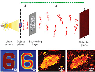
 Top: In scatter-plate microscope imaging, the imaging target is placed close to a thin scattering layer, and the aperture size is increased to achieve a high NA; the image is detected at a plane far from the scattering layer. Bottom: Comparison of conventional microscope images of a detail from a U.S. Air Force test target and a section of pine wood stem; in each pair, a conventional microscope image with monochromatic illumination is shown at the left and the corresponding scatter-plane microscope image is at the right.
For many centuries, lens quality has dominated microscope imaging—and, despite many efforts, no clear alternative to conventional lenses has emerged. High-resolution imaging exhibits particular demand for high-definition microscope objective lenses. As a result, prices of objective lenses have continually increased.
Traditionally, the properties of lens-based microscope objectives—fixed focal length, high numerical aperture (NA), short working distance, limited depth of focus (DOF) and magnification—have often set restrictions in their application. Aberration correction for microscope objectives requires an extended design consisting of a number of spherical lenses (sometimes more than 10), which increases complexity, bulkiness and price.
We have developed an alternative imaging system based on Freund’s intensity-correlation technique,1 which permits lensless imaging of an object using the memory effect in a scattering medium. 2 Specifically, we propose a new imaging device called a scatter-plate microscope, in which a conventional objective lens is replaced by a simple scatter plate.3 The approach can, our experiments suggest, turn an essentially costless scattering surface into a high-resolution microscope objective.
The scatter-plate microscope has several unique characteristics that we believe set it apart from traditional microscopes. It is extremely thin and light, suitable for low-cost production and scalable in device size. It also has variable focal length and magnification, and is self-adaptive to any conjugate image planes. It has variable NA and field of view (FOV), and flexible, extensible working distances. It is compatible with reflection- and transmission-mode imaging and should be immune to phase disturbances and aberrations.
Thus, a single low-cost scattering plate can perform the tasks of various microscope objectives with different magnifications, NAs and focal lengths. A relatively wider FOV and variable DOF are exclusive features of the technique. Our cross-correlation-based approach, combined with a reference point source, is capable of imaging complex objects; we were able to easily obtain a resolution of 2.19 µm. And we believe that our work in visible wavelengths also opens up the possibility of using scattering media as imaging lenses for X-ray and ultraviolet wavelengths.
Researchers
Alok Kumar Singh, Giancarlo Pedrini and Wolfgang Osten, Universität Stuttgart, Germany
Mitsuo Takeda, Utsunomiya University, Japan
References
1. I. Freund. Physica A 168, 49 (1990).
2. A.K. Singh et al. Light Sci. Appl. 6, e16219 (2017).
3. A.K. Singh et al. Sci. Rep. 7, 10687 (2017).