
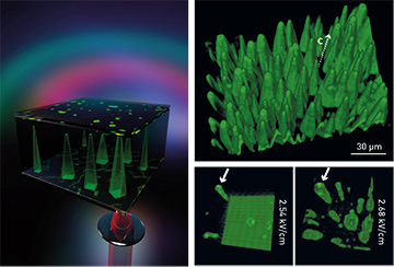
 Left: Sketch of 3-D Čerenkov SHG microscopy. A tightly focused infrared fs laser pulse is scanned through the crystal volume, and noncollinear Čerenkov SHG is generated at the domain walls. Right: 3-D image of micro-scaled needlelike ferroelectric domains in strontium barium niobate (top); 3-D images showing growing domains at two time points (bottom).
Ferroelectrics are pivotal materials widely used in sensor applications, data storage, and optical processing.1 These materials are characterized by the presence of ferroelectric domains: 3-D electric dipoles of the same polarization, separated by the so-called domain wall. Although ferroelectric materials have been widely employed and studied in the past, the spatio-temporal dynamical phenomena, including domain emergence, growth and switching, still prompt active debate.
Numerous techniques exist to examine and visualize ferroelectric domains, including chemical etching, piezo force microscopy and Raman spectroscopy. These methods, however, either are invasive, destroying the material, are limited to the crystal surface, or can measure only steady-state characteristics. Although researchers demonstrated seven years ago that Čerenkov-type second-harmonic generation (SHG) microscopy could image ferroelectric domains in 3-D,2 this advantageous technique has, surprisingly, rarely been applied since then, and has not yet been fully exploited.
In recent work, we have applied and extended Čerenkov-type SHG microscopy for in situ 3-D imaging of the switching process of ferroelectric domains, and have demonstrated the technique for the relaxor-type ferroelectric crystal strontium barium niobate (SBN).3 We have combined the optical method with the application of an external electric field, to record the current that flows during the dynamics of domain-wall growth.
The technique can optically detect lateral and longitudinal domain-wall motion with sub-micrometer resolution in the bulk starting from the walls’ nucleation. A comparison with the material’s hysteresis loop reveals the connection between the domain kinetics and the ferroelectric aging effect in SBN caused by the impurities and vacancies. In one significant outcome, we measured the pronounced growing-space direction of the domains in bulk material in 3-D, and established that the domains grow faster in the longitudinal than in the lateral direction. Our study has also revealed the process leading to the individual domain shape.4
Our results offer insights into one of the most complex processes in ferroelectricity. With these findings, the domain formation process can be explored in real-time and in 3-D as a function of many parameters, such as frequency of the electric field, temperature,5 or light illumination, all of which are essential for ferroelectric domain engineering.
Researchers
Jörg Imbrock, Mousa Ayoub, Cornelia Denz and Hannes Futterlieb, Westfälische Wilhelms–Universität Münster, Germany
References
1. P. Ferraro et al. Ferroelectric Crystals for Photonic Applications (Springer, 2009).
2. Y. Sheng et al. Opt. Express 18, 16539 (2010).
3. M. Ayoub et al. Adv. Mater. 29, 1603325 (2017).
4. M. Ayoub et al. Opt. Lett. 36, 4371 (2011).
5. M. Ayoub et al. Opt. Mater. Express 7, 3448 (2017).