
Optics Innovations
MUSE: Slide-Free Microscopy
Alexandre Y. Fong and Richard M. Levenson introduce a new optics-enabled microscopy method that creates diagnostic-quality images without lengthy and expensive specimen and slide preparation.
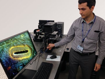 Farzad Fereidouni, assistant project scientist at U.C. Davis Medical Center, focuses the MUSE benchtop prototype to image a pig kidney artery. [U.C. Davis]
MUSE Microscopy Inc., a recent start-up from the University of California Davis (U.C. Davis) and Lawrence Livermore National Laboratory, USA, has developed a new, slide-free approach to microscopy. MUSE, which stands for microscopy with ultraviolet surface excitation, is a relatively simple, quick and inexpensive histological imaging method that can provide enhanced, diagnostic-quality images from whole tissue specimens.
…Log in or become a member to view the full text of this article.
This article may be available for purchase via the search at Optica Publishing Group.
Optica Members get the full text of Optics & Photonics News, plus a variety of other member benefits.