
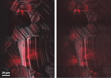
 Multimodal diffraction-limited (left) and super-resolved MP-SPIFI (right) images of CdTe solar cells, with nonlinear photoluminescence (red) and second-harmonic generation (gray) measured simultaneously.
In recent years, a menagerie of super-resolution imaging methods has emerged—techniques that provide spatial resolution well below the diffraction limit, offering powerful new information about the behavior of biological specimens on fine spatial scales. Most super-resolution microscopy thus far, however, has exploited the photophysics of fluorescent molecules. And, though several techniques for super-resolution fluorescence microscopy have found wide use in routine imaging of cells, no approaches have emerged that demonstrate super-resolution imaging in scattering tissues.
Recently, we introduced a super-resolution-imaging method, multiphoton spatial-frequency-modulated imaging (MP-SPIFI), that provides enhanced resolution for both luminescent and coherent nonlinear interactions. Using that technique, we demonstrated super-resolved imaging of two-photon excited fluorescence and second-harmonic generation.1
MP-SPIFI builds on previous work that forms images by illuminating an object with a periodic spatial illumination and collecting light on a single-pixel detector. This forms projections of the object’s spatial-frequency information onto the spatial frequency of the illumination light. The spatial frequency of the illumination pattern is then swept linearly in time, so that the temporal signal encodes the spatial frequency content of the object.2,3
In MP-SPIFI, we illuminate the object with three femtosecond laser pulses focused into light sheets. The crossing angle of two of the light sheets varies in time, sweeping the spatial frequency in the beam interference. The intense femtosecond light sheets drive a nonlinear excitation in the object. For a nonlinear order of η, MP-SPIFI can provide resolution up to 2η times the diffraction limit.
The image formation in MP-SPIFI is impervious to scattering between light emission and collection on a single pixel detector. Consequentially, MP-SPIFI opens a path for super-resolution imaging deep inside of scattering tissues. Moreover, the scaling of the resolution enhancement with nonlinear interaction order means that exploring higher-order nonlinearities will enable even larger enhancements in spatial resolution—for example, by exploiting three-photon fluorescence with long-wavelength femtosecond laser pulses.4 This provides a path to super-resolved imaging at unprecedented depths in highly scattering media.
Researchers
Jeffrey J. Field, Keith A. Wernsing and Randy A. Bartels, Colorado State University, Fort Collins, Colo., USA
Jeff A. Squier, Colorado School of Mines, Golden, Colo., USA
References
1. J.J. Field et al. Proc. Natl. Acad. Sci. USA 113, 6605 (2016).
2. G. Futia et al. Opt. Express 19, 1626 (2011).
3. J.J. Field et al. Optica 3, 971 (2016).
4. N.G. Horton et al. Nat. Photon. 7, 205 (2013).