
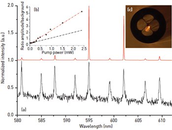
 (a) WGM spectra of a 10-μm-diameter polystyrene microsphere attached to the tip of a MOF below (black) and above (red) its lasing threshold. (b) The lasing characteristic of the microsphere. (c) A lasing microsphere attached to a fiber in which the propagation of WGMs in the equatorial plane can be clearly seen. [François et al. Sensors 15, 1168 (2015)].
Whispering gallery mode (WGM) spectroscopy has been at the forefront of developments in optical biosensing. This is largely due to WGM’s exceptional molecular-level detection limits.1 However, using WGM spectroscopy outside of research environments remains a considerable challenge because WGMs, whether in spheres, toroids or capillaries, are interrogated using a cumbersome phase-matching coupling scheme with either a fiber taper or a prism. Any fluctuation of the gap between the resonator and the taper or prism induces a variation in the resonance positions, which is detrimental to biosensing applications in which the spectral shift of the resonances is used to infer changes in the surrounding refractive index.
Fluorescent microresonators, which permit remote excitation and readout of a WGM-modulated fluorescence signal, do not suffer from the same practical constraints and have therefore attracted considerable attention for in vivo sensing applications.2 However, the lower Q factors realized for fluorescent resonators, typically orders of magnitude below their passive resonator counterparts, pose a significant limitation.
Our recent work demonstrates that by combining the unique light-guiding properties of microstructured optical fibers (MOF) with fluorescent microspheres, some of the intrinsic limitations of fluorescent microresonators can be alleviated. Modelling WGMs in fluorescent microspheres has allowed us to pinpoint the ideal diameter of microsphere resonators for a large range of materials, enabling optimization of both the refractive index sensitivity and resolution, and hence, detection limit.3 We have also shown lasing in what we believe is the smallest resonators ever reported in aqueous solution (Ø = 10 μm polystyrene) by tuning the dye concentration to avoid dye self-quenching and consequently minimizing the lasing threshold.4 Moreover, achieving WGM lasing of a microsphere at the tip of a MOF results in unprecedented Q factor enhancements—for example, 2 × 10⁴ compared to about 103 for the same free-floating microsphere below its lasing threshold.
Building on these results, we have shown that this simple approach of combining lasing microspheres with MOFs improves the detection limit of specific biomolecules when used as a dip sensor, thereby paving a pathway for new in vivo biological sensing modalities.5
Researchers
Alexandre François, Shahraam Afshar V. and Tanya M. Monro, University of Adelaide, Australia; University of South Australia
Tess Reynolds, Nicolas Riesen, Jonathan M.M. Hall and Matthew R. Henderson, University of Adelaide
References
1. Baaske et al. Nat. Nanotech. 9, 933 (2014).
2. Humar et al. Nat. Photon. 9, 572 (2015).
3. Reynolds et al. Opt. Express 23, 14784 (2015).
4. François et al. Appl. Phys. Lett. 106, 031104 (2015).
5. François et al. Sensors 15, 1168 (2015).