
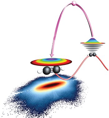
 Mechanism of LIED. A broadband electron wave packet is created by ionization of C2H2 at the peak of an intense mid-IR field. After accelerating and gaining energy in the optical field, the wave packet can return as a plane wave at a later time and diffract off its parent ion. Accurate information about both the C–H and C–C bond lengths can be extracted from the momentum distribution of the diffracted electrons.
Francis Crick, the co-discoverer of the double-helix DNA structure, reportedly said “If you want to understand function, study structure.” This basic principle underlies the development of structural studies in biology, chemistry and physics. The mysteries linked to the structure of the DNA molecule and the biological functions that it undergoes are now solved due to diffraction and microscopy-based imaging techniques. However, these biological functions are instigated by much more rapid phenomena that are still not well understood. To image such mechanisms, a technique that can achieve both sub-angstrom spatial and few-femtosecond temporal resolutions is required.
Laser-induced electron diffraction (LIED) is a strong-field ionization- based imaging methodology, and it is currently the only technique that is able to achieve the abovementioned resolution requirements. So far, structure determination efforts using LIED have been limited to diatomic molecules, which do not possess the many degrees of freedom found in molecules relevant to chemistry and biology. This is due to the demanding experimental capabilities that must both create high-energy core-penetrating electrons able to probe the molecular structure, and capture all of the correlated products related to the strong-field induced-diffraction process.
Using a unique combination of a 160-kHz mid-IR optical parametric chirped pulse amplification (OPCPA) source with a reaction microscope (REMI) charged-particle detection system, we have developed an experimental recipe that solves both of the above critical issues and permits the investigation of polyatomic molecules using LIED.1,2 We demonstrated the potential of our methodology by accurately imaging the complete structure of the organic molecule acetylene (C2H2), including the C–H bond that is considered highly elusive in conventional diffraction imaging techniques. Additionally, we showed how to harness the intrinsic temporal properties of strong-field ionization to image molecular structure at unprecedented, fewfemtosecond resolutions.
Our results provide the foundation for dynamical investigations of larger molecular systems and set the scene for the first “molecular movie” with simultaneous picometer and few-femtosecond resolutions. Using the roadmap outlined in our work, the ultrafast phenomena that prompt biological functions may soon be probed on their natural spatial and temporal scales.
Researchers
M.G. Pullen, B. Wolter, M. Baudisch and J. Biegert, ICFO–Institut de Ciencies Fotoniques, Barcelona, Spain
A.-T. Le and C.D. Lin, Kansas State University, Manhattan, Kan., USA
M. Hemmer, DESY–Deutsches Elektronen Synchrotron, Hamburg, Germany
A. Senftleben, Universität Kassel, Kassel, Germany
C.D. Schröter and R. Moshammer, Max-Planck-Institut für Kernphysik, Heidelberg, Germany
J. Ullrich, Physikalisch-Technische Bundesanstalt, Braunschweig, Germany
References
1. M.G. Pullen et al. Nat. Commun. 6, 7262 (2015).
2. B. Wolter et al. Phys. Rev. X, 5, 021034 (2015).