
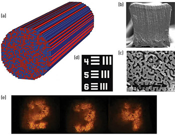
 (a) Sketch of the dielectric medium. (b) Cross-section of polymer Anderson-localized fiber, with an approximate side width of 250 µm. (c) SEM image of a 24 µm wide region on the tip of the fiber (darker regions have lower refractive index). (d) A section of the 1951 U.S. Air Force resolution test chart used in imaging tests. (e) Images transported through a segment of the disordered fiber.
In pioneering work in 1989, Hans De Raedt and colleagues studied an optical wave system in which the refractive-index profile is random in the transverse plane and invariant in the longitudinal direction.1 They showed that an optical beam launched in the longitudinal direction can become localized in the transverse plane, due to the strong random scattering from the transverse random index fluctuations, and can propagate in the longitudinal direction with a finite beam radius, as with a conventional optical fiber. They attributed this behavior to a 2-D manifestation of Anderson localization,2 occurring in the transverse plane of the beam. This phenomenon, which they referred to as transverse Anderson localization, was observed experimentally in 2007 in a photo-refractive crystal utilizing refractive-index variations on the order of 0.0001.3
In 2012, our team demonstrated transverse Anderson localization of light in a polymer random optical-fiber medium with refractive-index fluctuations of order 0.1.4 Such large fluctuations are essential for device applications if the mean localized beam radius is to be comparable with or smaller than that of conventional optical fibers. Our polymer Anderson-localized fiber allowed for simultaneous propagation of multiple beams in a single strand of disordered optical fiber, with potential applications in beam-multiplexed optical communications and optical imaging.
This year, we successfully demonstrated that the polymer Anderson-localized fiber can be used for endoscopic fiber optic imaging.5 The earlier demonstration of spatial beam multiplexing suggested the possibility of using disordered optical fiber for some form of image transport; our 2014 work, however, also showed that the image transport can have comparable or higher quality than some of the best commercially available multicore imaging optical fibers, with less pixelation and higher contrast. Remarkably, the high-quality image transport is achieved because of, not in spite of, the imaging system’s high level of randomness.
Future efforts will concentrate on reducing the attenuation of the disordered fiber for longer-distance image transport, and on developing an air-glass disordered fiber to achieve a smaller beam localization radius for higher image transport resolution.
Researchers
Salman Karbasi, University of California, San Diego, Calif., USA
Ryan J. Frazier, University of California, Berkeley, Calif., USA
Karl W. Koch, Corning Inc., Corning, N.Y., USA
Thomas Hawkins and John Ballato, Clemson University, Clemson, S.C., USA
Arash Mafi, University of New Mexico, Albuquerque, N.M., USA
References
1. H. De Raedt et al. Phys. Rev. Lett. 62, 47 (1989).
2. P.W. Anderson. Phys. Rev. 109, 1492 (1958).
3. T. Schwartz et al. Nature 446, 52 (2007).
4. S. Karbasi et al. Opt. Lett. 37, 2304 (2012).
5. S. Karbasi et al. Nat. Commun. 5, 3362 (2014).