
Scatterings
Transient Absorption Microscopy Images Nanotubes in Cells
Researchers at Purdue University have learned how to detect both metallic and semiconducting nanotubes within biological tissue.
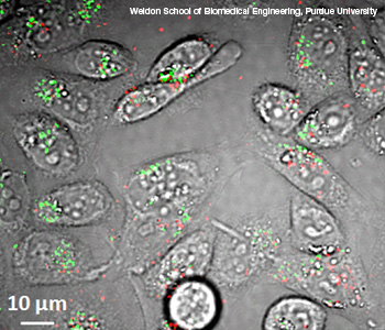 False-color transient absorption micrograph shows both metallic (red) and semiconducting (green) single-walled carbon nanotubes in live hamster cells.
Weldon School of Biomedical Engineering, Purdue University
Single-walled carbon nanotubes have unique optical properties that may hinder imaging them in vivo. Researchers at Purdue University (U.S.A.) have learned how to detect both metallic and semiconducting nanotubes within biological tissue (Nat. Nanotechnol., doi:10.1038/nnano.2011.210).
…Log in or become a member to view the full text of this article.
This article may be available for purchase via the search at Optica Publishing Group.
Optica Members get the full text of Optics & Photonics News, plus a variety of other member benefits.