
Scatterings
Multi-Mode Imaging Probe Could Detect Ovarian Cancer
Researchers have developed a multi-mode imaging probe that could examine ovarian tissue via minimally invasive surgery.
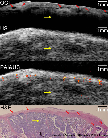 Co-registered images of malignant ovarian tissue obtained with the hybrid imaging device. (Top to bottom) OCT image, ultrasound image, superimposed photoacoustic and ultrasound image and corresponding histology. Yellow arrow: malignant tissue.
Ovarian cancer is difficult to diagnose in its early stages because symptoms often don’t manifest themselves until the disease has spread, and also because there is no effective screening method. Researchers at the University of Connecticut and the University of Southern California (U.S.A.) have developed a multi-mode imaging probe that could examine ovarian tissue via minimally invasive surgery (Biomed. Opt. Exp. 2, 2551; doi: 10.1364/BOE.2.002551).
…Log in or become a member to view the full text of this article.
This article may be available for purchase via the search at Optica Publishing Group.
Optica Members get the full text of Optics & Photonics News, plus a variety of other member benefits.