
Scatterings
Imaging Drug Effects on Tumors
Holographic tissue dynamics spectroscopy allows researchers to look at motion inside living tissue.
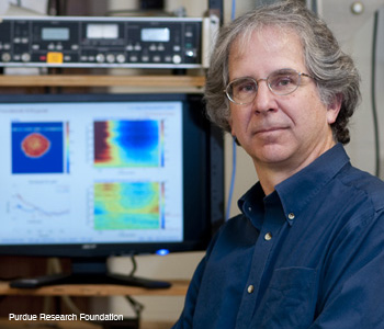 David Nolte with spectrograms created using holographic tissue dynamics spectroscopy. The spectrograms show how cells react to drugs.
Holographic tissue dynamics spectroscopy—a method developed by Purdue researcher David Nolte with colleagues at Purdue—allows researchers to look at motion inside living tissue. It could provide a way to detect the effects of new drugs on cancerous tumors. At the recent LabAutomation Conference in Palm Springs, Calif., U.S.A., Nolte says, "It generated a lot of excitement because it is a new form of high-content screening that works without labels that could be valuable in drug discovery." (See "Motility Contrast Imaging in Three-Dimensional Tissue-Based Drug Screening," by D. Nolte et al., LabAutomation2011.)
…Log in or become a member to view the full text of this article.
This article may be available for purchase via the search at Optica Publishing Group.
Optica Members get the full text of Optics & Photonics News, plus a variety of other member benefits.