
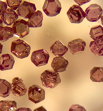
A new multimodal-imaging system uses microdiamonds, around 200 μm across, as tracers to enable simultaneous optical and NMR imaging. [Image: Ashok Ajoy, UC Berkeley]
For imaging biological tissues, clinicians often want sharp images of features at great tissue depths. Modern imaging technologies, though, force a choice: optical wavelengths cannot penetrate far without scattering, and nuclear magnetic resonance (NMR), which relies on radio frequencies as well as magnetic fields, goes deep without detail.
A team of scientists at a U.S. university has explored a new method of combining the two imaging modes that could yield better results than either technique alone (Proc. Natl. Acad. Sci. USA., doi: 10.1073/pnas.2023579118). The key: seeding the target tissue with specially designed microparticles of diamond that show up in both types of imaging.
Tiny diamonds with flaws
The researchers at the University of California Berkeley, USA, employed 200-μm-wide tracer particles made of specially flawed diamond. Although pure diamond consists solely of carbon atoms, these microparticles contain a couple of strategic flaws.
First, nitrogen atoms, paired with a lattice vacancy, replace some of the carbon atoms in the crystalline lattice of the diamond. These point defects—called nitrogen–vacancy centers, and a staple in studies of quantum technology—fluoresce brightly when laser light hits them. Also, these particular diamond particles had enriched levels of the spin-polarized 13C isotope, which shows up better in NMR imaging than the dominant 12C isotope.
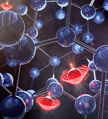
Nitrogen–vacancy centers in microdiamonds can be polarized (red spinning balls) and made to emit red light when illuminated by a laser. The polarized centers then hyperpolarize nearby 13C atoms (blue balls), allowing them to be detected by NMR imaging. This allows the tracers to be imaged both by optical fluorescence microscopy and NMR, providing higher resolution pictures deeper inside tissue, according to the researchers. [Image: Xudong Lv and Mustafa Kamran, UC Berkeley]
According to the researchers, this combination of fluorescent centers and spin-polarized carbon atoms allows the diamonds, acting as tracers, to be imaged at the same time both by optical fluorescence microscopy and NMR.
Experimental results
For a baseline test, the Berkeley team, led by chemistry professor Ashok Ajoy, created a phantom made of a 4.2-mm-wide ring of diamond microparticles and imaged it separately with green laser light and NMR equipment. As expected, the test ring gave off red fluorescence and the magnetic resonance image looked fuzzy. Then, the researchers tested the dual-mode imaging of the phantom under a variety of conditions, including the addition of dyes and methanol, with and without background suppression techniques.
In a press release accompanying the work, Ajoy characterized the work as “perhaps the first demonstration that the same object can be imaged in optics and hyperpolarized MRI simultaneously”—two modes that when used in combination “are better than the sum of their parts.” The scientists report that the dual-mode technique works at room temperature and a comparatively low magnetic field of roughly 40 mT. Extension of the technique to in vivo work depends on whether it meets safety limits for the specific absorption rates of all types of radiation involved—and whether a living body can eliminate the diamond particles.