
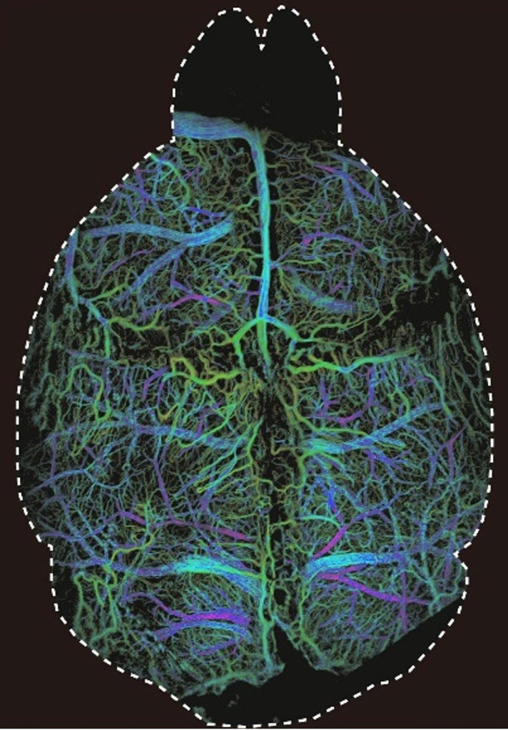
An image taken with the new DOLI method shows microcirculation in a mouse’s brain in 3D. [Image: ETH Zurich, University of Zurich / Daniel Razansky] [Enlarge image]
The brain’s workings are a long-standing medical mystery. For nearly a hundred years, scientists have developed ways to peer beneath the skull—but with existing methods it’s still difficult to see the tiny, complex blood flows in living brain tissue.
A new technique aims to change that. Called diffuse optical localization imaging (DOLI), the technique has reportedly doubled the depth at which fluorescence microscopy can noninvasively probe the brain, allowing researchers to see blood flows in mouse brains for the first time without having to remove the skull (Optica, doi: 10.1364/OPTICA.420378).
Simple setup, big results
Fluorescence microscopy uses fluorescent contrast agents that are released into the bloodstream. When irradiated at the right wavelength, tiny particles in the agent emit a glow that can be captured on camera to reveal the complex networks of the circulatory system in the brain.
Previously, scattering and absorption by tissue between the glowing particles and the camera resulted in fuzzy images. But using a specific spectral region—the second near-infrared window, from 1000 to 1700 nm—can significantly reduce these scattering and absorption effects. By pairing a new lead sulfide–based quantum dot contrast agent, which operates in the wavelength window, with ultra-efficient shortwave infrared cameras, the researchers were able to noninvasively image 3D capillary-level blood flow in mice for the first time.
“The most exciting aspect of the new DOLI technique is the ease of implementation,” said co-author Daniel Razansky, a professor of biomedical imaging at ETH Zurich and the University of Zurich. “You basically need a relatively simple and affordable camera setup. No expensive pulsed lasers or sophisticated optics needed.”
Eyeing extension to human studies
The technique was first tested on synthetic tissue models that showed clearly resolved images at depths up to four millimeters. Additional tests on live mice demonstrated the technique’s noninvasive, high-resolution cerebrovascular imaging capabilities. With a one-centimeter field of view—large enough to cover a mouse’s brain—the technique was able to pinpoint individual droplets and visualize blood flow speeds. By measuring the apparent size of the droplets, the researchers were also able to determine their depth in the brain, allowing for 3D imaging.
The researchers hope that the technique could someday be used for studying neurological disorders, which are thought to affect a billion people. However, additional advances will be required first, given the size differences between human and mouse brains.
“It is of paramount importance to develop new noninvasive imaging techniques to be able to better understand how the brain works, [and] determine the underlying biological causes of neurodegenerative and other brain diseases,” Razasnsky said.