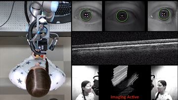
Duke University’s new optical coherence tomography scanner is attached to a robotic arm that can move the scanner to align with a patient’s eyes. [Image: Duke University] [Enlarge image]
Biomedical engineers and ophthalmologists from Duke University, USA, have designed and demonstrated a hands-free automated version of a standard desktop optical coherence tomography (OCT) scanner. The research team believes that the setup may expand the valuable technology’s reach to primary care offices and emergency departments.
In proof-of-concept demonstrations, the scanner, which is attached to a robotic arm, produced images of the cornea and retina that are comparable to standard tabletop systems—in less than 10 seconds (Nat. Biomed. Eng., doi: 10.1038/s41551-021-00753-6).
Conquering tremor and patient movement
OCT is a 3D imaging technology used to get micron-scale images of anatomic structures within the eye. The technology has revolutionized the diagnosis and monitoring of eye diseases, including age-related macular degeneration, diabetic retinopathy and glaucoma. Unfortunately, OCT scanning is often limited to specialist clinics, because the tabletop devices are bulky and require a trained ophthalmic photographer for operation.
By automating the entire imaging process and bringing the scanner to the patient, the Duke researchers hope to make regular OCT-based eye exams accessible to everyone. The automated OCT workspace includes two 3D cameras to the left and right of a robotic arm. The 3D cameras help position the OCT scanner mounted on the robotic arm in front of the eye. Once the robotic arm is in the general vicinity of the eye, more sensitive cameras on the arm precisely line up the OCT scanner with the pupil. If the patient moves their head, the robotic arm uses active tracking technology to keep the scanner aligned with the pupil.
According to lead author Mark Draelos, “The robotic arm gives us the flexibility of handheld OCT scanners, but we don’t need to worry about any operator tremor. If a person moves, the robot moves with it. As long as the scanner is aligned within a centimeter of where it needs to be on your pupil, [it] can get an image that is as good as a tabletop scanner.” The scan for each eye is completed in less than 10 seconds, with the entire alignment, scan, and imaging process taking about 50 seconds.
Proof-of-concept demonstrations
The Duke researchers designed active-tracking anterior (corneal) and retinal scan heads for proof-of-concept demonstrations. First, they characterized image quality for each of the OCT scan heads using optical simulations. Next, they measured the active-tracking ability of the scan heads with phantoms and with free-standing individuals.
Both scan heads produced images that were sufficient for examining the cornea and retina. Both heads also demonstrated active-tracking accuracy of phantoms and free-standing individuals that “satisfied” their aiming needs, with residual motion artifacts corrected in post-scan processing.
Finally, the researchers eliminated the operator entirely and tested fully autonomous OCT corneal and retinal imaging with five free-standing individuals with healthy eyes. Results were again positive, with clear views of the individuals’ corneas and retinas. An expert reviewer assessed the quality of the images from the fully automated scans and determined that the Duke scanner’s performance is comparable to current tabletop OCT systems.
Limitations and next steps
While the demonstrations and expert’s review are promising, the researchers recognize that their autonomous OCT scanning system’s biggest limitations are image resolution and, for retinal scans specifically, small field-of-view. The team is now focused on refining the scanner, and see implementing the technology in primary care offices and emergency departments as “the logical next step.”

