
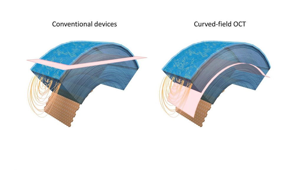
Traditional imaging approaches acquire a flat slice (pink) that crosses through several corneal layers at once, limiting the field of view (left). The curved-field OCT approach matches the curvature of the cornea to provide a larger imaging area (right). [Image: Viacheslav Mazlin, The Langevin Institute] [Enlarge image]
Improved diagnosis and treatment of some eye diseases and other health conditions could result from an optical coherence tomography (OCT) instrument that can image curved layers of corneal tissue, reports a research team in France (Optica, doi: 10.1364/OPTICA.396949).
Dubbed curved-field OCT (CF-OCT), the new instrument marries full-field OCT (FF-OCT) with a principle of classical Twyman-Green interferometer design to allow for the first time successively wider, planar views of the cornea, including curved portions that follow the natural curvature of the tissue atop the eyeball, the researchers say.
A new possibility
“The developed CF-OCT brings the new possibility of cell-resolution corneal imaging capturing optical sections of arbitrary curvature,” says project leader Viacheslav Mazlin of the Langevin Institute in Paris and CNRS, the French national research agency. Other team members include researchers at Sorbonne University and Quinze-Vingts National Eye Hospital, both in Paris; and the University of Pittsburgh, USA.
The technique works, Mazlin says, by embedding into the FF-OCT design a curved rather than flat mirror—in this case, a curved lens with enough reflectivity to act as a mirror.
“The main breakthrough lies in the understanding that the curved interference field—well known from the classical Twyman-Green interferometer—can be useful not only for imaging the surface shape, but also for making an optical sectioning to see the internal curved layers,” Mazlin says. “Technologically it is the combination of an optical sectioning full-field interferometer with a curved lens that is new.”
With CF-OCT, Mazlin says, “we can curve the interference field not only for comparing surface shapes in both arms of the interferometer, but for optical sectioning of internal sample layers with the curved field.” He says the latter is particularly interesting for imaging samples with curved layers like the cornea that must not be physically flattened. He says CF-OCT, which the team was able to test in vivo, offers a 10-times-wider viewing area than the clinical state of the art.
For now, the device employs a joystick and motorized stages for manual focusing during imaging, but future iterations may provide autofocus capabilities, Mazlin says.
A choice of lenses
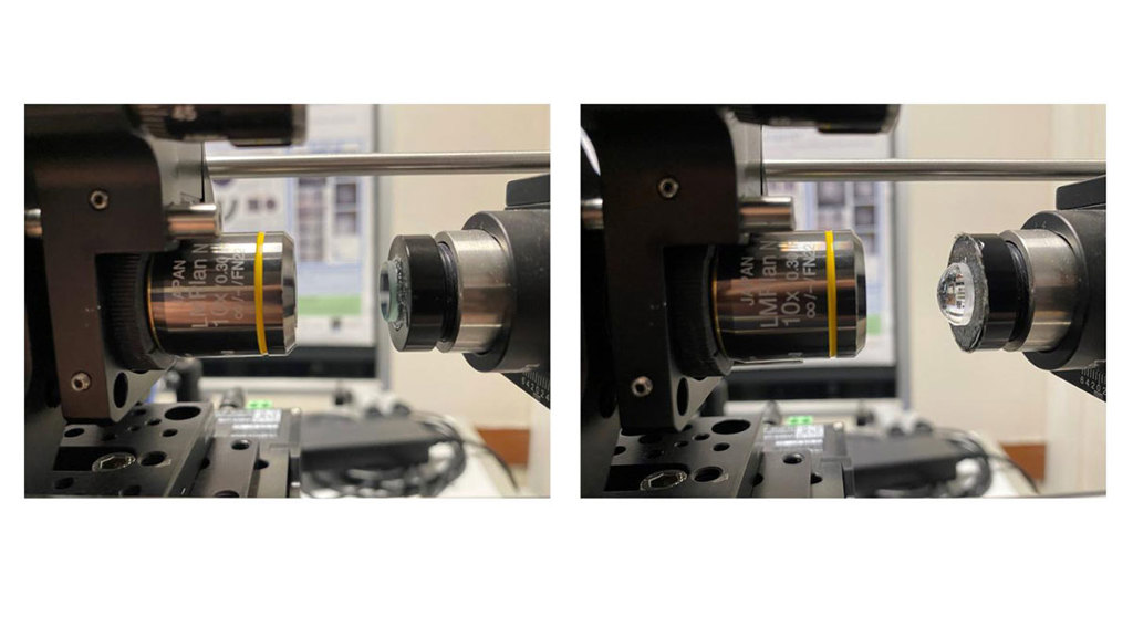
To capture images from a curved sample, the researchers replaced the flat mirror (left) of a full-field OCT setup with a curved optical lens (right). The curved lens optically flattens the acquired images. [Image: Viacheslav Mazlin, The Langevin Institute] [Enlarge image]
“We use two different lenses to achieve the best results in imaging the anterior and posterior parts of the cornea, which naturally have different curvatures,” Mazlin says. “We switch the curvature of optical sectioning by manually replacing the lenses and realigning the device. The realigning procedure takes about half an hour, which is too long to be used in the clinical setting.”
However, “in the article we show a solution to this problem when the curvatures are not very different from each other, as is the case with the cornea,” Mazlin says. The instrument’s four-phase tomographic image retrieval scheme can be used to suppress dense fringe pattern artifacts that result from the differing tissue curvatures, he says, and reveal the mosaic from the posterior portion of the cornea, even when only the lens matching the curvature of the anterior portion is used.
“If the curvatures to be switched are very different—for example, concave cornea to convex retina—then tunable lenses can potentially be embedded into the interferometer to control the curvature,” Mazlin says. “The latter approach potentially enables automatic, fast curvature correction.”
Cell-level resolution
Mazlin says the new instrument provides cell-level resolution not possible with conventional OCT—which has just tens of micrometers resolution—nor practical with a confocal microscope, which requires contact with the eye and has a small viewing area below 0.2 mm2. If CF-OCT can be further developed for the clinic, he says, it could help to come up with better disease monitoring and treatment strategies for a variety of conditions involving the cornea and general health, such as diabetes, which is known to alter corneal nerve density.
Such imaging capabilities, he says, could also be used to improve outcomes for corneal transplants and cataract surgery with dramatically improved precision of endothelial cell counting before and after surgery. Potentially the device could be used during eye surgery, Mazlin says. However, “We see the first step for the novel instrument is imaging prior to surgery and surgical follow up.”
But CF-OCT would be useful for imaging all kinds of transparent curved objects, which should not be flattened, Mazlin says. In conventional imaging methods, he says, curved sections can be reconstructed from 3D volumetric images, however, this requires long acquisition time.
“The benefit of CF-OCT is that the curved optical section is obtained directly in two fast camera shots using a simple instrumentation, and the time-consuming 3D imaging is not needed,” he says.
Toward a clinical device
The device concept is still several steps away from becoming a clinical product, Mazlin says. But he says the instrument will next be tested in patient cohorts, exhibiting various corneal, ocular and general health disorders.
“In the future, CF-OCT would benefit from incorporation into conventional OCT, enabling an automatic aligning procedure and, potentially, combining a large view of the entire anterior eye segment with cell resolution,” Mazlin says. The addition of tunable lenses may enable easy switching between CF-OCT to FF-OCT configurations, with the latter being beneficial for extending functionality of the device with angiography and retinal imaging.