
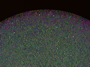
Detail of cross-section of multimode bundle containing some 30,000 closely spaced optical fibers. Australian scientists have used the bundle to create ultra-slim light field imaging probes. [Image: Anthony Orth, RMIT University]
Medical and optical scientists are racing to develop all-optical biopsy techniques for faster and less invasive cancer diagnoses (see “Toward an All-Optical Biopsy,” OPN, April 2019). Endoscopes powered by flexible bundles of optical fibers already exist for peering inside the human body, but the images they produce are limited to two dimensions, usually because of size constraints on the instrument.
Researchers at an Australian university have learned how to extract depth information from the light path of the fiber bundles in today's clinical microendoscopes (Sci. Advances, doi: 10.1126/sciadv.aav1555). The technique produces 3–D images that could give a better view of potential tumors during optical biopsies.
The team at RMIT University in Melbourne, Australia, took its inspiration from light-field imaging systems, which focus light rays computationally rather than mechanically. Current microendoscopes contain bundles of multimode optical fibers, which scramble the light field while propagating it.
Intracore insight
![]()
Anthony Orth, a research fellow at RMIT University in Melbourne, Australia, holds an ultrathin microendoscope used in the study. [Image: RMIT University]
Anthony Orth, an RMIT biophotonics research fellow, and his colleagues realized that the light output from these fiber bundles still contains intracore intensity information about the light input to the bundle. (A typical endoscopy setup suppresses the intensity data to reduce pixelation.) Using a bundle containing roughly 30,000 fibers with an average center-to-center spacing of 3.2 μm, the scientists calculated the angular distribution of 550-nm light coming into the bundle from a point source much smaller than the bundle’s cross-section.
According to the researchers, the fibers at a greater angle of incidence from the center of the bundle have more excited higher-order modes than the fibers near the center. The farther the individual fibers are from the bundle center, the more the light rays are pushed to the edge of the individual fiber core. Next, the scientists processed the light with a technique called digital aperture filtering, then mathematically modeled the light field to realize a focused image.
The RMIT light field technique is relatively insensitive to moderate bends in the fiber bundle—always a major concern when inserting a long, thin imaging device into the human body. The team cautions, however, that the device won't work when tissue is fundamentally unobservable due to light attenuation.